|
|
Case Reports Indian Pediatrics 2002; 39:1152-1156 |
||
|
Relapsing Hypertrophic Osteoarthropathy in a Child with Bronchiectasis |
||
|
Figen Özcay
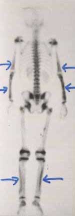
Hypertrophic osteoarthropathy (HOA) is knwon to occur in patients with chronic suppurative pulmonary infections and bronchiectasis(1). HOA is a rare condition characterized by clubbing, periostosis of the tubular bones, and arthritis-like signs and symptoms, first described in association with intrathoracic inflammatory lesions in 1889-90(2). Hypertrophic osteoarthropathy is common in adults, particularly in individuals with pulmonary malignancies. In children, the disease has been noted in various conditions associated with chronic pulmonary infection, such as cystic fibrosis, immunodeficiency syndromes, immotile cilia syndrome, inflammatory bowel diseases, and cyanotic congenital heart disease(3). In this report, we describe an adolescent boy with bronchiectasis whose arthritic signs and symptoms flared during acute episodes of underlying chronic pulmonary infection. He was investigated for the presence of septic and rheumotoid arthritis, systemic lumpus erythematosis and amyloidosis several times. To the best of our knowledge, relapsing arthritis responding to antibiotics with each bout of pneumonia in bronchiectatic children with HOA has not been described in the English literature before. Case Report A 14-year-old boy was admitted to our hospital with a 2-week history of cough, fever, shortness of breath, and painful hands, feet and legs. The child had been hospitalized many times for pneumonia since he was 2 years old. He was diagnosed with bronchiectasis secondary to recurrent pulmonary infection at 5 years of age, and clubbing was noted at that time. Swelling and pain in his hands and feet were occurring off and on for last 4 years and used to be aggravated with each bout of pulmonary infection. The boy had already been investigated for possible causes of bronchiectasis, acute and chronic arthritis, particularly amyloidosis, prior to his presentation at our hospital. Physical examination revealed that the patient was 142 cm tall (below the 3rd percentile) and weighed 32 kg (in the 10th percentile). His axillary temperature was 38.5ºC and his respiratory rate was 46/min. He had halitosis, his antero-posterior chest diameter was larger than normal, and he exhibited dyspnea, intercostal retractions, and bilateral crackles and rhonchi on lung ausculation. We also noted clubbing of the toes and fingers. His ankles, wrists, and the lower parts of his legs were warm, swollen, and painful to touch. Laboratory studies revealed the following results: hemoglobin 11g/dL; white blood cell count 8.4 × 10⁹/L with 72% neutrophils on a peripheral blood smear; calcium 9.2 mg/dL; phosphorus 3.3 mg/dL; alkaline phosphatase 513 U/L; erythrocyte sedimentation rate 75 mm/h; and C-reactive protein 59 mg/L. The levels of alpha-1 antitrypsin, IgA, IgG, IgM, IgE, and complement C3 and C4 were all normal. Serologic studies for anti-neutrohil cytoplasmic antibodies (c-ANCA and p-ANCA), rheumatoid factor, anti-nuclear antibody, and anti-DNA antibody were all negative. The arterial blood pH was 7.46, PaCO2 was 38 mmHg, and PaO2 was 73 mmHg. Arterial oxygen saturation, as determined by a pulse oxymeter, was 89%. Repeated quantitative sweat tests were normal for several times since he was 2 years old (8,16,21 and 17 mEq/L). A chest X-ray showed extensive pulmonary infiltration, honeycombing and cystic changes with airfluid levels. High resolution computerized tomography of the thorax revealed thickening and irregularity of the bronchial walls, bronchial dilation, and alveolo-interstitial infiltration, all of which are consistent with bronchiectasis. Radiographs of the extremities demonstrated symmetrical new bone formation (periosteal reaction) involving the distal portions of the tibia and fibula bilaterally. Bone scintigraphy showed an intense and symmetrical uptake up to 99mTc-MDP in the distal parts of the humerus, radius, and ulna bilaterally, and a linear increase in uptake at the cortical margins of the tibia and femur in both legs (Fig 1). These scintigraphic and radiological findings were consistent with HOA. Hemophilus influenzae was cultured from the patient’s sputum, but mycobacterial and fungal cultures of sputum samples were negative. Electron microscopic examination of the nasal mucosa showed no structural abnormalities of the ciliated epithelial cells. There was no amyloid deposition detected in a rectal biopsy specimen. The patient was prescribed a 3-week course of intravenous antibiotics (ceftazidime, amikacin, and metronidazole). By the end of treatment, the pain and heat in his extremities had resolved completely and the swelling had decreased. Repeated bone scintigraphy showed minimal regression of the previously mentioned findings in his bones.
Discussion Patients with HOA usually seek medical attention because of the severe pain and swelling in their joints and extremities. Radiographs of the long bones reveal periosteal reaction, and bone scans show increased uptake of 99mTc-MDP along the periosteal surfaces of the affected bones. Our patient exhibited the typical features of HOA, and we suspected that the condition was related to his bronchiectasis and chronic pulmonary infection. His medical history included episodes of increased joint pain and swelling concomitant with high fever, production of purulent sputum, and cough. The latter three symptoms all suggest that he had pulmonary infection during these episodes. Also, although his arthritis-like symptoms regressed when he responded to the treatment for pneumonia, his ankles and wrists remained thicker than normal and the clubbing persisted. Reversal of HOA through cure of the underlying condition has been reported in patients who have had malignancies removed, and in liver disease patients who have undergone liver transplantation(4). If HOA does not resolve, this may indicate poor prognosis in cases of malignancy, or graft rejection in transplant recipients. Resolution of the disease have also been reported after treatment in patients with AIDS who have Pneumocystis carinii pneumonia and in patients with tuberculosis(5,6). Although our patient’s clinical signs and symptoms of joint disease did subside after aggressive antibiotic therapy, we suspect that the severity and persistence of his chronic pulmonary inflammation had already produced permanent radiological and scintigraphic findigs of HOA. The exact mechanism and pathogenesis of clubbing and HOA is unknown. Research has shown that low arterial oxygen content alone does not cuase the condition. Although there is a significant correlation between clubbing and arterial hypoxemia, results from a series of children with a variety of lung diseases indicated that clubbing was present in only 46% of those who exhibited arterial oxygen desaturation(7). In 1987, Dickinson and Martin postulated that there was a "clubbing-producing particulate substance" that, in normal individuals, is removed from the blood by the lung(8). They suggested that the normal pulmonary vascular bed retains these large particles, but that a right-to-left shunt, which is seen mainly in CCHD, would allow them to bypass the lung. More recent invstigations have demonstrated that this "particulate material" is actually composed of megakaryocytes, platelet clumps, and individual platelets. Platelets and megakaryocytes escape from the lung capillaries, travel to the distal parts of the extremities, and ultimately reach the nail beds. The platelets and megakaryocytes contain and release platelet-derived growth factor, which is general growth promoter and mitogen. This factor increases vascular permeability, and has a chemotactic effect on monocytes and neutrophils. It is also a powerful attractant for smooth muscle cells and fibroblasts. The overall effect leads to clubbing and HOA. Other growth factors produced by megakaryocytes and platelets, such as vascular endothelial growth factor(9), hepatocyte growth factor, and transforming growth factor beta-1, have also been implicated. The pathogenesis of HOA is also obscure in the specific setting of chronic pulmonary infection and bronchiectasis. These patients develop clubbing, but is not necessarily associated with arterial oxygen desaturation. Vascular dilatation in the lung tissue surrounding a pyogenic infection may facilitate the escape of platelet clumps from the pulmonary capillaries. Also, all inflammatory conditions are associated with platelet aggregation and clot formation. In bronchiectasis, marked hypertrophy of the bronchial vasculature may be associated with the development of systemic-pulmonary shunts, which might help explain the occurrence of HOA in these patients(10). Bronchiectasis is still common and constitutes a health care problem in developing countries(11). Any pediatrician who works in these area have a good chance to encounter a child with bronchiectasis and HOA. Besides, bronchiectasis is known to occur in association with rheumatoid arthritis, and with Sjögren’s syndrome and amyloidosis, which are also characterized by arthritis. HOA should always be included in the differential diagnosis for joint disease in patients with bronchiectasis. Contributors: FO, NO and US were involved in the care of the patient, conceiving the article, drafting of manuscript and final approval. Funding: None. Competing interests: None stated.
| ||
|
References | ||
|
![]()