 |
Case Reports Indian Pediatrics 1999;36: 1270-1272 |
Radiological Demonstration of Ascaris in Esophagus |
Dheeraj Gandhi, Deep N. Srivastava, Bobby Batra* Satish Chandra From the Departments of Radiodiagnosis and Pediatrics*, All
India Institute of Medical Sciences, Ansari Nagar, New Delhi 110 029, India. |
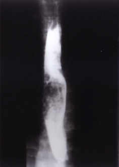
Ascaris lumbricoides is a cosmopolitan parasite inhabiting the gut of one fourth of world's population. The highest prevalence is in malnourished people residing in the developing countries(1) and jejunum and ileum are its preferred habitats. However, demons-tration of these worms in esophagus is extremely unusual. We report one such case. Case Report A 15-year-old male child presented with two months history of recurrent abdominal pain, fever and easy fatiguability. The child was having retrosternal burning sensation and dysphagia for solid foods for a day before presentation. The child was of lean built weighing 28 Kg with a height of 130 cm. Positive findings on physical examination included severe pallor, koilonychia and several enlarged cervical jugular lymph nodes measuring 0.5-0.8 cm in diameter. The lymph nodes were mobile and not matted. Systemic examination was unremarkable. Laboratory examination was within reference range except for a hemoglobin count of 6.2 g/dl, hypochromic microcytic peripheral smear and elevated ESR (32 mm, 1st hour, Wintrobe). Mantoux test yielded a reading of 5�5 mm. Chest X-ray and an ultrasound examination of abdomen was normal. Fine needle aspiration cytology from the cervical lymph nodes had revealed reactive inflam-matory cells. Barium study for the evaluation of dysphagia revealed multiple mottled and linear filling defects expanding the middle one-third of thoracic esophagus (Fig. 1). There was no evidence of esophageal mucosal irregulariy, abnormal stasis, disordered motility or any distal esophageal strictrure.
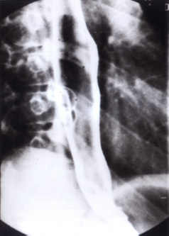
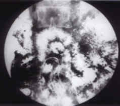
Fig. 1. Barium swallow demonstrating mottled filling defects expanding the esophageal lumen. A diagnosis of esophageal bezoar was entertained on the basis of radiological appear-ance and the patient was scheduled for endoscopic removal of bezoar. However, later in the day, the patient vomited out a bunch of six live Ascaris lumbricoides worms and felt symptomatically better subsequently. We repeated the barium upper GI series the next day to look for any residual worms in the esophagus or their distal migration into the stomach. Repeat barium examination revealed normal eso- phageal lumen (Fig. 2) and mucosa. Delayed fluoroscopy and spot films (Fig. 3) documented the presence of numerous worms in the small intestine and ascending colon.
Fig. 2. Esophageal lumen and mucosa are normal on the barium swallow done the next day.
Fig. 3. Barium meal follow through reveals a large number of worms in the small intestine. Discussion Ascaris infestation is particularly common in India. Although a vast majority of worms are found in jejunum and ileum, numerous complications due to abnormal migration of worms can occur such as billiary and pancreatic duct blockages and obstruction of appendix(2). Adult worms can also be vomited out through the nose causing great distress to the patient(3), and a particularly revolting sight. Demonstration of the roundworms in the esophagus is distinctly unusual. Tomas et al. have reported esophageal and stomach Ascaris as an exceptional endoscopic observation(4). Baird et al. have reported massive Ascaris infestation in a two year old child. At autopsy, their patient had roundworms in the esophagus, stomach, bile ducts, gall bladder and small intestine(5). Our search of literature (1966 onwards) produced only two cases where roundworms have been visualized in the esophagus on barium swallow examina-tion(6,7). The roundworms were seen as tubular filling defects in the esophageal lumen in both the illustrations. In our patient, however, the roundworms in the esophagus gave an appearance of mottled filling defect with the expansion of esophageal lumen. The radiological appearance was not unlike that of bezoars, leading to misdiagnosis. Therefore, this possibility must be borne in mind when one comes across such a radiographic appearance in esophagus. A short history of dysphagia and rapid transit of worms across the esophagus in our patient suggests that we demonstrated the worms in the esophagus during their abnormal anti-peristaltic migration. References 1. Koncman EW, Allen SD, Janda WM, Schreckenberger PC, Winn Jr WC. Color Atlas and Textbook of Diagnostic Microbiology, 5th edn, Philadelphia, Lippncot, 1997; p 1100. 2. Srivastava DN, Chakravarti AL, Saxena R, Saraswat V, Kulshreshtha A, Gujral B, et al. Sonographic appraisal of endobiliary ascaris with calculi. Am J Gastorenterol 1991; 86: 527-528. 3. Howard BJ. Clinical and Pathogenic Microbiology, 2nd edn. St Louis, Mosby Year Book Inc, 1994; p 669. 4. Tomas A, Vida F, Puig X. Presence of Ascaris lumbricoides in esophagus and stomach: An exceptional endoscopic observation. Gastro-enterol Hepatol (spanish) 1997; 20: 335. 5. Baird JK, Mistrey M, Pimsler M, Connor DH. Fatal human ascariasis following secondary massive infection. Am J Trop Med Hyg 1986; 35: 314-318. 6. Reeder MM, Palmer PES. Radiology of Tropical Diseases. Baltimore, Williams and Wilkins Company, 1981, pp 411-438. 7. Reeder MM. The radiological and ultrasound evaluation of Ascariasis of gastrointestinal, biliary and respiratory tracts. Semin Roentgenol 1998, XXXIII: 57-78. |