|
|
Case Reports Indian Pediatrics 2000;37: 901-903 |
||
|
Spontaneous Neonatal Esophageal Perforation |
||
From the Referral Neonatal Unit, Department of Pediatrics, and Department of Pediatric Surgery*, Maulana Azad Medical College, New Delhi 110 002, India. Reprint requests: Dr. N.B. Mathur, Professor of Pediatrics, Maulana Azad Medical College, New Delhi 110 002, India. Manuscript Received: August 10, 1999;
Spontaneous esophageal perforation or ‘Neonatal Boerhaave’s Syndrome’ is a rare catastrophe(1). Esophageal perforation may be fatal and delayed treatment causes an increase in morbidity and mortality(2,3). Since its first clear description(4), the trend has been towards non operative management in the neonate(5,6). We report one such case.
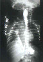
A 2-day-old female baby born at home by spontaneous vaginal delivery at term was admitted with the complaints of respiratory distress for one day and an episode of bluish discoloration of the body once 24 hours back. At 24 hours of life, the baby choked after a feed at night and turned blue, but the color improved following physical stimulation by the mother. Following this episode, excessive salivation was noticed. At admission, the baby was slightly dusky, with respiratory distress and pooling of secretions in the mouth. Orogastic catheter could be negotiated easily. On investigating, the sepsis parameters were not suggestive of infection. Chest X-ray revealed a right sided pneumothorax with underlying collapse. A tube thoracostomy was done. Nasogastric tube feeds were started on day three but she became dusky and hence was kept nil by mouth. Repeat sepsis screen was positive for infection. Later, nasogastric tube feeds were restarted but the condition of the baby deteriorated again. Thoracostomy revealed frank pus. Pus culture was sterile. On day 7, there was suspicion of milk in the chest tube and the possibility of an esophageal rupture was entertained. A gastrograffin swallow study was performed which revealed a dilated upper thoracic eso-phagus and a normal lower thoracic esophagus. Mediastinal tracking of contrast into the right pleural cavity was seen arising from the upper esophagus at D3 level (Fig. 1). The baby was managed conservatively and she started improving slowly. A repeat gastro-graffin swallow study on day 17 of admission showed a persistent leak though the size had decreased. A repeat contrast study was done at one month of admission which showed no evidence of leakage.
Esophageal rupture is labeled as sponta-neous perforation, Boerhaave’s syndrome, effort perforation and esophageal apoplexy if no instrumentation or intubation of the eso-phagus has occurred, implying that an intrinsic anatomic abnormality or dysfunction is present within the esophageal wall(7-9). The causes of esophageal perforation in the neonate can be spontaneous, instrumental (following esophagoscopy, dilatation and intubation), traumatic or anastomotic. Sponta-neous perforation of the esophagus in neonates and infants is rare, accounting for only 4% of all reported cases(10). Noniatrogenic perfora-tions are extremely rare and these are usually seen in full term infants. The most common site is the lower end of esophagus. Etiological hypotheses for spontaneous perforation vary, and include increased abdominal pressure at delivery, perinatal hypoxemia, peptic esophagitis and gastroesophageal reflux (GER)(7,8,9,11). Iatrogenic perforation of the esophagus is more commonly seen in premature, small for gestational age infants(12-14) and usually occurs in the cervical esophagus. Pharyngeal suctioning with a stiff catheter, endotracheal intubation and traumatic laryngoscopy have all been described as etiological factors(15). The usual clinical history in neonatal Boerhaave’s syndrome includes dyspnea followed by cyanosis which may occur shortly after birth but is usually delayed for six to thirty hours. Respiratory distress is caused by pneumothorax that has prediliction for the right side (as opposed to left side in the adults). This is explained by the close adherance of the aorta to the left side of the esophagus in the infant which provides additional support to that side. Vomiting, coughing, choking episodes and mild hematemesis have been observed in several cases. Thoracocentesis or tube thoracostomy may reveal serosanguinous or grossly bloody pleural fluid or the contents of previous feedings(11,16). In contrast, esophageal perforation is most frequently associated with difficulty in passing a nasogastric tube in premature infants (in 63% of cases)(17,18). Subcutaneous emphysema is rare in infants unless massive pneumomediastinum occurs. There are a number of differences between spontaneous neonatal esophageal rupture and those occurring in adults. In adults, the hydropneumothorax is not under tension(19), mediastinal emphysema is present in approxi-mately 60%(20), males predominate and rupture usually occurs into the left pleural cavity(20). In the neonate, tension penumo-thorax or tension hydropneumothorax is a characteristic feature, pneumomediastinum is uncommon, females predominate and rupture usually occurs into the right pleural cavity(1). Chest radiograph alone may be diagnostic of esophageal perforation when a hydro-pneumothorax is seen combined with air dissecting in the mediastinum. An unusual course of the nasogastric tube may be the first indication of this diagnosis. In all cases, esophagography is necessary to establish the diagnosis, localize the perforation and direct therapy. It should always be performed under fluoroscopic control. The classical appearance is of "double esophagus". Esophagoscopy offers no diagnostic advantage and may actually enlarge the perforation. Routine surgical intervention does not appear to improve the rate of survival in these critically ill newborns. Tube thoracostomy must be placed. If there is clinical deterioration or respiratory compromise, and closed chest tube drainage does not handle the leak, direct repair of the perforation is indicated. The overall mortality from pharyngeal or esophageal perforation in newborns is approximately 30%. | ||
|
|