|
|
Case Reports Indian Pediatrics 2000;37: 893-896 |
||
|
Langerhans Cell Histiocytosis Involving the Urinary Bladder |
||
From the Departments of Child Health, Pathology # and Radiodiagnosis*, Christian Medical College Hospital, Vellore, Tamilnadu, India; and ## Department of Oncology, Krishna Nursing Home, MG Road, Ernakulam, Kerala, India. Reprint requests: Dr. Thomas Cherian, Professor of Child Health, Christian Medical College Hospital, Vellore 632 004, Tamilnadu, India. E-mail: cheri@cmcvellore.ac.in Manuscript Received: October 11, 1999; Langerhans cell histiocytosis (LCH) is a disease characterized by proliferation and infiltration of various organs with pathological Langerhans cells. The organs commonly involved are bone, skin, lymph nodes, bone marrow, liver, spleen, lungs and endocrine system. The rarer sites include thymus, gastrointestinal system, female genital tract and urinary tract(1). LCH involving the urinary bladder is very rare. A medline search over 20 years has not revealed any cases of LCH affecting the urinary bladder. We report a case of LCH with urinary bladder involvement in an 11-year-old boy.
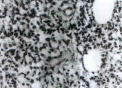
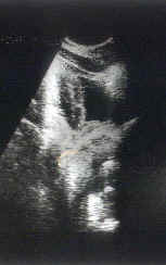
An 11-year-old boy presented with swelling of the left maxilla, cervical lymphadenopathy and hepatosplenomegaly in July 1997. This was diagnosed as LCH based on histopathological examination. He was treated with etoposide, vincristine and prednisolone for five months, following, which his symptoms resolved. He remained well till February 1998 when he developed hematuria. A cystoscopic examina-tion and fine needle aspiration was performed, which was consistent with LCH. Chemotherapy was restarted in April 1998 using etoposide, vinblastine and prednisolone weekly till July 1998. However, he continued to have hematuria and also developed recurrence of left maxillary swelling. Hence he was referred to our hospital for further management. On physical examination, his weight was 30 kilograms and height was 131 cm. His vital signs were normal. There was no pallor. He had enlarged upper deep cervical lymph nodes bilaterally (2 ´ 2 cm each). There was a non-tender swelling over the left maxilla, measuring 5 ´ 5 cm, with consistency varying from firm to hard. There were no other bony swellings. The liver was palpable 2 cm below the right costal margin. There was no splenomegaly. Examination of other systems did not reveal any abnormality. Investigations revealed hemoglobin of 10 g/dl, total leukocyte count of 26,400/cu mm with neutrophils 38%, eosinophils 17%, lymphocytes 39%, and monocytes 5%. Platelet count was 3,51,000/cu mm. Liver function test showed a total bilirubin of 0.5 mg/dl, direct bilirubin 0.1 mg/dl, total protein 7.8 g/dl, albumin 4.4 g/dl, SGOT 19U/L, SGPT 11U/L, and alkaline phosphatase 160 U/L. Urine microscopy revealed 4-6 WBC and 40-45 RBC per high power field. There were no casts or crystals. Bone marrow examination was normal. Bone scan showed increased tracer uptake in the left maxilla. A skeletal survey of other bones was normal. The biopsy slides of the maxillary swelling and urinary bladder were reviewed, which were consistent with LCH (Fig. 1). The cells were S-100 positive on immunochemistry. An ultrasound of the abdomen showed an area of irregular thicken-ing on the wall of the urinary bladder, measuring 4.7 ´ 2.5 cm (Fig. 2). He was diagnosed to have LCH with multisystem involvement in view of the maxillary swelling and urinary bladder involvement. It was decided to start him on LCH-1 protocol, plan A. He was given methyl-prednisolone (30 mg/kg) intravenously once daily for three days followed by vinblastine (6 mg/m2) on the fourth day. Hematuria persisted despite chemotherapy but the maxillary swelling resolved. He was then given radiotherapy to the urinary bladder, 1600 cGy in eight fractions using 60 Co teletherapy unit. Following this his symptoms subsided. In September 1999, eleven months after completion of treatment he was asymptomatic and repeat cystoscopy did not show any residual lesion.
LCH is a rare disease of childhood with protean manifestations. It can present at any age, from neonatal period to old age. But the disease is more common in children with a peak incidence from one to three years. Males are more commonly affected than females(2). The etiology and pathogenesis of LCH remains unknown. It is currently thought to be a clonal proliferative disorder with highly variable biologic behavior and clinical severity(3,4). The disease is self-limited in some, whereas in some others it is extensive with multi-organ dysfunction. LCH affects many organs in the body with varying frequency. Involvement of the urinary tract is rare. Tsuchiya et al. reported a case of peri-renal mass of LCH in a 15-month-old girl(5). There are two other reports of renal eosinophilic granuloma in adults(6,7). A case of eosinophilic granuloma of the ureter was also documented(8). To our knowledge, urinary bladder involvement in LCH has not been documented before. We are reporting this case to document the involvement of the urinary bladder in LCH. Contributors: LGM and TC were involved in preparation of the manuscript and care of the patient. SN reported the biopsy and performed immunohisto-chemistry RAC provided radiological assistance. MNCN was involved in treatment of the patient. TC will act as the guarantor for the paper.
| ||
|
Key Messages |
||
|
||
|
|