Pulmonary alveolar microlithiasis (PAM) is
characterized by the formation of calcium microlith in the alveoli due
to defective clearance of phosphates. The clinical traits of PAM are
heterogeneous and lung deterioration progresses at different speeds even
when microliths appear early. The hallmark of PAM is clinico-radiological
dissociation with typical imaging findings that correlate with specific
pathological findings [1]. This report highlights the clinico-radiological
dissociation and the tests available for the diagnosis of PAM.
Three children were diagnosed as PAM between 2015 and
2019. The first case was a 5-year-old girl (14.6 kg) who presented with
complaints of fever, cough and poor appetite for seven months. Physical
examination was normal. Imaging showed lung infiltrates suggestive of
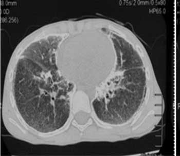
interstitial lung disease (Fig. 1 and 2) but the
open lung wedge biopsy showed calcific nature of the lesions confirming
the diagnosis (Web Fig. 1). The second one was a
4½-year-old girl (16.3 kg) who presented with recurrent fever, poor
appetite and cough. The radiological imaging showed micronodular
mottling on both lung fields. Broncho-alveolar lavage (BAL) demonstrated
pus cells with moderate Streptococci. Video assisted thoracoscopic lung
biopsy demonstrated air-spaces with innumerable tiny calcified bodies
that are concentrically laminated with radial striations in the intra
alveolar lumen consistent with pulmonary alveolar microlithiasis. The
third case was a 12-year-old girl (17.3 kg) who presented with fever,
weight loss and poor appetite for one year. She was treated for
tuberculosis. Imaging showed calcified micronodule lesions on the
midzones of both the lungs and ground glass opacities. Pulmonary
function testing showed a mild restrictive pattern. BAL demonstrated
Moraxella spp.
 |

|
|
Fig. 1 Chest radiograph showing
diffuse reticulonodular opacities in both lung fields.
|
Fig. 2 Computed tomography lung window
showing calcified micronodule lesions on the midzones with
ground glass opacities.
|
PAM is reported worldwide with more than half of the
cases from five countries (Turkey, China, Japan, India, and Italy) [2].
It is often diagnosed incidentally during radiography of the chest [3].
All the three children in the present study were girls, contrary to a
study from Turkey which reported six boys with PAM and a familial
inheritance [4]. Studies have identified mutations causing decreased
cellular uptake of phosphate leading to formation of intra-alveolar
microliths [5].
The clinico-radiological dissociation with
significant radiological findings in the absence of lower respiratory
features like dyspnea and retractions have been reported earlier [6]. As
the evolution of PAM is insidious, diffuse micronodular opacities may
appear as miliary shadows in the chest radiograph leading to
misdiagnosis of tuberculosis. CT chest demonstrated multiple calcified
micronodules in both lungs, subpleural regions, and in the cardiac
margins in all three affected children. PAM is usually diagnosed on the
basis of a typical radiological pattern like a sand-like micronodulation
of calcific density diffusely involving both lungs with basal
predominance. Presence of this pattern may preclude the need for a lung
biopsy. SLC34A2 is implicated as the defective gene [5]. Paucity
of symptoms and clinico-radiological dissociation may invite unnecessary
investigations in the initial stages of the disease when PAM should be
kept as a close differential.
1. Ferreira Francisco FA, Pereira e Silva JL,
Hochhegger B, Zanetti G, Marchiori E, et al. Pulmonary alveolar
micro-lithiasis. Respir Med. 2013;107:1-9.
2. Castellana G, Gentile M, Castellana R, Resta O.
Pulmonary alveolar microlithiasis: Review of the 1022 cases reported
worldwide. Eur Respi Rev. 2015;24:607-20.
3. Jönsson ÅLM, SimonsenU, Hilberg O, Bendstrup E.
Pulmonary alveolar microlithiasis: Two case reports and review of the
literature. Eur Resp Rev. 2012;21: 249-56.
4. Senyigit A, Yaramis A, Gürkan F, Kirbas G,
Büyükbayram H, Nazaroglu H, et al. Pulmonary alveolar
microlithiasis: A rare familial inheritance with a report of six cases
in a family. Respiration. 2001;68:204-9.
5. Huqun, Izumi S, Miyazawa H, Ishii K, Uchiyama B,
Ishida, et al. Mutations in the SLC34A2 gene are associated with
pulmonary alveolar microlithiasis. Am J Respir Crit Care Med.
2007;175:263-8.
6. Shah M, Joshi JM. Bone scintigraphy in pulmonary alveolar
microlithiasis. Indian J Chest Dis Allied Sci. 2011; 53:221-3.

