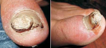
A 13-year-old boy presented with a ‘growth’ beneath the nail
of the right great toe (Fig. 1). The lesion
was painful and had been present for the preceding 6 months.
The nodule was tender, bony-hard in consistency, and
measured 20 x 15 mm in diameter. The nail plate showed
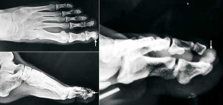
onycholysis. A radiograph revealed a calcified projection on
the dorso-lateral part of the distal phalanx, continuous
with the underlying bone (Fig. 2). Based on the
clinical presentation and radiological features, a diagnosis
of subungual exostosis was made.
 |
|
Fig. 1 Subungual nodule
with onycholysis.
|
 |
|
Fig. 2 Radiograph showing
calcified projection on the distal phalanx of the
great toe (white arrows).
|
Subungual exostosis is a relatively rare,
acquired, benign osteocartilaginous tumor occurring mainly
in children and young adults. They are found beneath the
distal edge of the nail, most commonly of the great toe.
However, other toes or, occasionally, a finger may be
involved. The first manifestation of this tumor is a
painful, small, pink or flesh-colored, hard, exophytic
growth that projects beyond the inner free edge of the nail.
The overlying nail becomes brittle and may be lifted or
become detached. The surface of the lesion may become
hyperkeratotic. The exact pathogenesis of exostosis remains
elusive. However, it probably reflects a reactive metaplasia
resulting from micro-trauma. It should be differentiated
from granuloma pyogenicum (sessile, friable, vascular
nodule, which bleeds easily on touch), verruca vulgaris (verrucous
nodule, devoid of skin markings; multiple bleeding points
are seen on pairing of the lesion), glomus tumor (skin-colored
or blue-red nodule; on palpation: extremely tender with
radiating pain), and squamous cell carcinoma (usually found
at the sulcus of the nail; presents as a growth under the
distal lateral edge of the nail; usually a long term history
of several years is present).
However, the bony consistency of the
nodule would usually suggest the correct diagnosis. Plain
radiography can generally confirm it, exhibiting an
exostotic tumor arising from the dorsal aspect of the distal
phalanx as in the present case. Complete excision or
curettage is the mainstay of treatment of this condition.


