| |
|
Correspondence Indian Pediatrics 2008; 45:704 |
|||
|
Klebsiella Septicemia Presenting as Renal Subcapsular Abscess and Hypertension in a Neonate |
|||
|
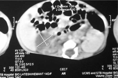
A term, appropriate for gestational age female baby weighing 3.0 kg was delivered uneventfully by vaginal route to a 28 year-old primigravida mother. The mother complained of fever and foul smelling vaginal discharge for 72 hours prior to delivery. Sepsis screen at birth revealed hemoglobin: 16 g/dL, TLC: 24,500 cells/mm3, platelet count: 1.8 lac/mm3, blood sugar: 34 mg/dL, and blood urea: 32 mg/dL. C-reactive protein was >6 mg/dL. On day 3, blood culture revealed Klebsiella pneumoniae, but urine and CSF cultures were sterile. Therapy with ceftriaxone and vancomycin were started. On day 8, hypertension was recorded in all four limbs, blood pressure readings being RUL: 82/69/52 mm Hg, LUL: 96/78/60 mm Hg, RLL: 100/90/69 mm Hg, and LLL: 96/78/60 mm Hg. On examination, there was no cyanosis or pallor, all peripheral pulses were well palpable, and liver was palpable 7 cm below the costal margin. Investigations revealed blood urea: 90 mg/dL, serum creatinine: 1.8 mg/dL, and a normal arterial blood gas. The baby was passing urine adequately. Intravenous furosemide and oral nifedipine were started. Ultrasonogram of abdomen revealed a hetrogenous collection adjacent to right kidney indenting the capsule. CT scan of the abdomen showed a subcapsular collection (2.7 × 1.9 × 1.5 cm) adjacent to the anterolateral part of the right kidney with no excretion of contrast in the right kidney even in delayed film (Fig. 1). Duplex scan of the renal vessels was normal. Blood urea and serum creatinine returned to normal by day 12 of life and hypertension resolved by day 15 of life. Baby was discharged after receiving parenteral antibiotics for 4 weeks. Follow-up CECT abdomen showed complete resolution of the subcapsular renal abscess at 3 months of age and prompt excretion of contrast into the pelvicalyceal system.
Renal and perinephric abscesses in the newborn are rare and are mostly acquired by hematogenous route, though ascending infections may occur via urethra(2). P-fimbriated Escherichia coli is the most frequent pathogen causing renal and peri-renal abscesses; Klebsiella pneumoinae is reported less frequently. Urine cultures are positive in only 10% of cases while blood cultures may be positive in up to 40% of cases(3,4).The mainstay of treatment for renal abscess is parenteral broad spectrum antibiotics, however abscesses larger than 3cm or those unresponsive to antibiotics may require percutaneous or open surgical drainage (5). Pooja Dewan and *Nitin Agarwal,
|
![]()