|
|
Letters to the Editor Indian Pediatrics 2006; 43:746-747 |
|||
|
Swyer-James Macleod Syndrome |
|||
|
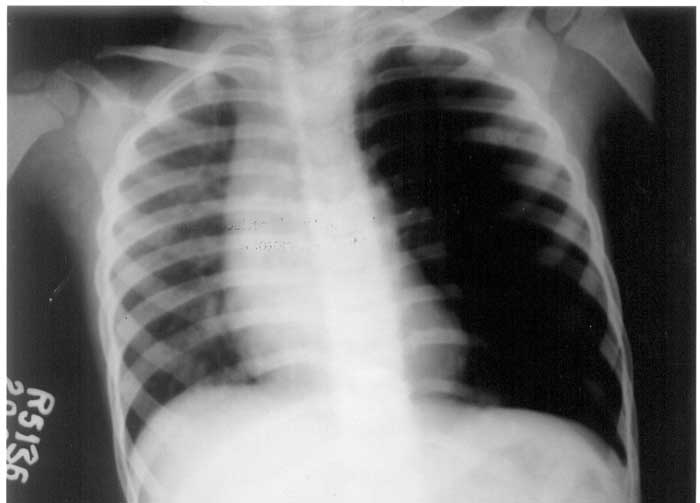
A two-year-old male child presented to us with chronic cough for past 6 months. The cough started following an acute respiratory illness of unknown etiology for which he had been hospitalized for two weeks. Examination revealed a well-preserved child with normal vital parameters and a SpO2 of 97% in room air. There was diminished breath sounds on the left side in the chest while rest of the physical examination including an ENT examination was unremarkable. Hemogram with ESR was normal. ELISA for HIV and Mantoux test were also negative. Chest radiograph revealed a hyperlucent lung with oligemic lung fields on the left (Fig.1). CT scan of the chest revealed similar findings and there was no evidence of any bronchiectasis. A lung perfusion scan with Tc99m labeled MAA showed markedly decreased tracer uptake in left lung with normal homogenous distribution on the right. Ventilation scan and pulmonary function test could not be carried out, as the child was very small to cooperate for the same. He obtained no benefit with a trial of bronchodilators, has been administered pneumococcal and influenza vaccines apart from routine immunization and is on regular follow up. Swyer and James first described the syndrome in 1953 followed by macleod in 1954(2). The disease starts as an obliterative bronchiolitis with concomitant vasculitis commonly following infections with organisms such as adenovirus, measles or pertussis. The damage to the terminal or respiratory bronchioles in early childhood possibly prevents normal development of their alveolar buds(1). One study of bronchoalveolar lavage in SJMS suggests an ongoing inflammatory process in the lung(3). The condition should be differentiated from congenital anomalies of airway/pulmonary vessels and bronchial obstruction due to mucus plug or foreign body(1). Management is based on (i) pulmonary function test, which may reveal mixed restrictive and obstructive airway disease (ii) prevention and/or follow up for infection and bronchiectasis and (iii) rarely surgery(4,5). B.M. John,
|
![]()