|
|
Images in Clinical Practice Indian Pediatrics 2006; 43:740 |
||||
|
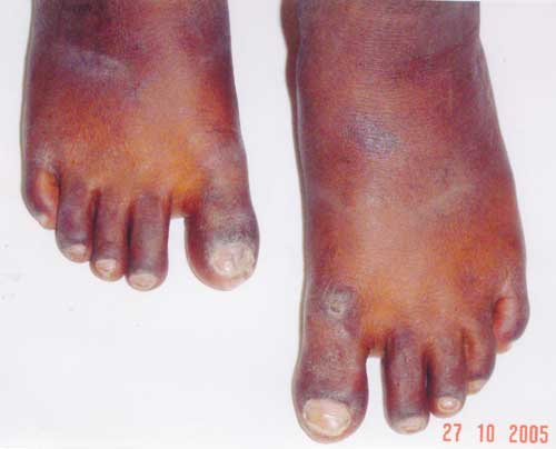
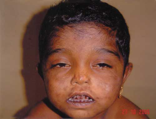
Congenital Erythropoeitic Porphyria |
||||
|
This autosomal recessive condition besides its typical skin, dental and urine findings can also have ocular and hematologic findings. Erythrodontia can also be seen in fluorosis, tetracycline therapy, food stains or dentinogenesis imperfecta. Porphyrial skin lesions must be differentiated from xeroderma pigmentosum, epidermolysis bullosa and pemphigoid. The best therapy is avoidance of sunlight, while oral beta-carotenes have been tried with limited benefit. A.N. Prasad, |
![]()