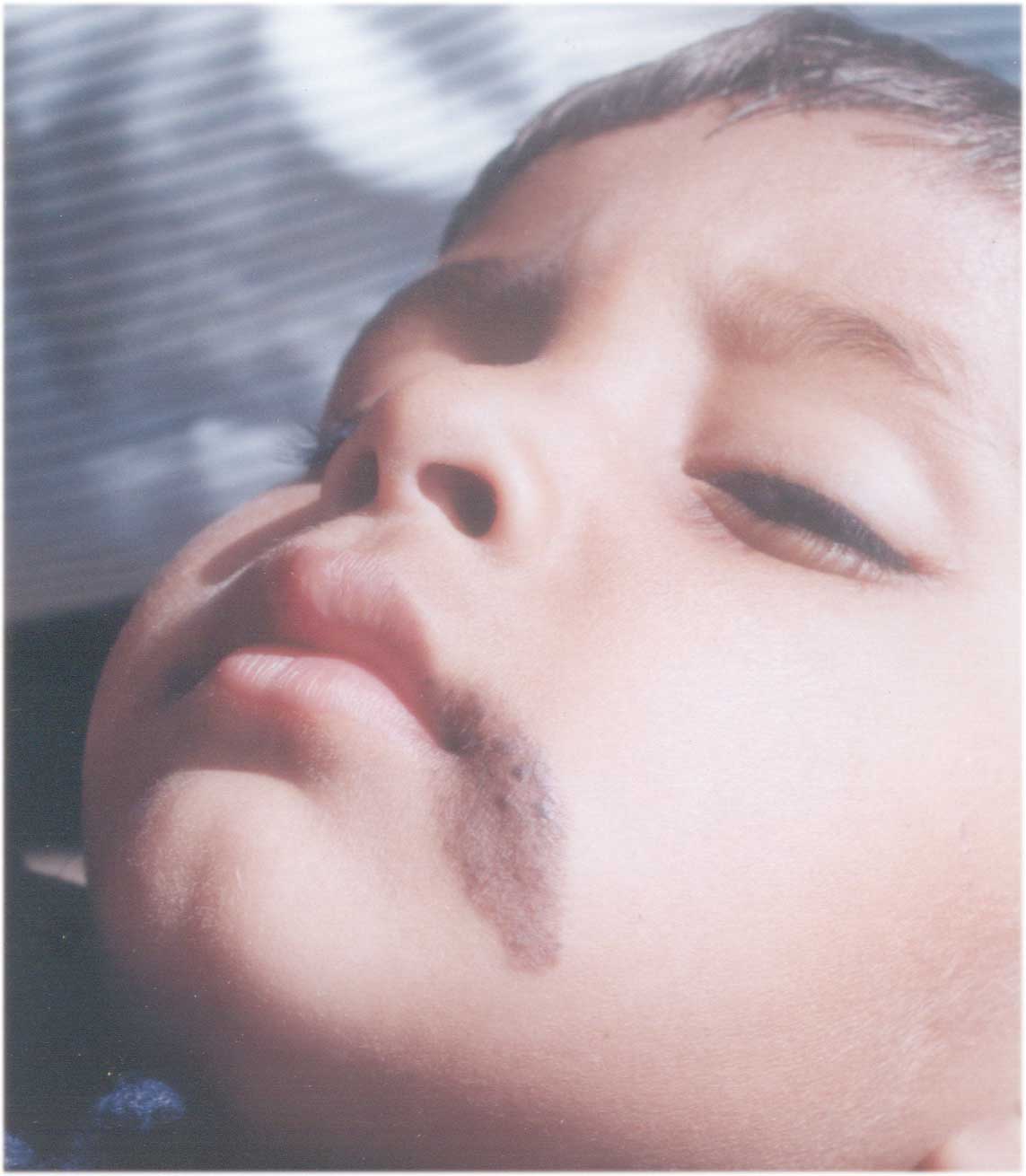
A four-year-old boy presented with an asymptomatic, well-defined, light
brown colored elliptical patch, 1.5 × 3.2 centimetres in size, on left
cheek adjoining the angle of lip (Fig. 1). The vermilion border
was involved while the oral mucosa was clear. Superimposed on the
homogeneous light brown tan, small darker-brown papular lesions were
evident, as if sprinkled over the underlying base. Rest of the body was
free of any pigmented lesion. History revealed presence of the
underlying homogeneous lesion since birth, while the superimposed
papular lesions had started appearing from last two years, and were
still evolving. Histopathology from the lightly colored base was
consistent with that of a cafe au lait macule, while from a
darker papule was suggestive of a compound melanocytic nevus.
 |
|
Fig. 1. Nevus Spilus Comprising of well-defined
homogeneous café au lait macule with darker overlying compound
melanocytic nevi. |
Nevus Spilus (Speckled Lentiginous Nevus) is
described as a hyperpigmented patch with superimposed darker macules
and/or papules. Surrounded by ‘congenital versus acquired’ controversy,
the lesions are benign and usually start in infancy. Hypothesized to be
due to a field defect in melanoblasts, genetic and environmental factors
also seem to playa role. Considering a small chance of malignant
transformation, observation and serial photographs remain the mainstay
of management. Surgical excision may be considered in some cases.
Ram Gulati,
Consultant Dermatologist,
C-118, Shivajai Marg,
Tilak Nagar,
Jaipur 302 204, India.
E-mail: [email protected]
