|
|
Images in Clinical Practice Indian Pediatrics 2004; 41:854-855 |
||||
|
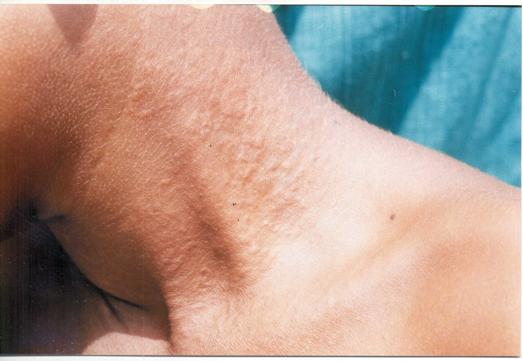
Yellow Skin Papules Over the Neck-Pseudoxanthoma Elasticum |
||||
|
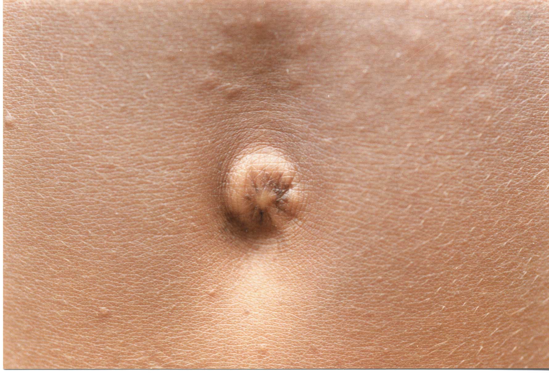
On examination, multiple, discrete as well as coalescent, skin colored to yellowish, indurated, papular lesions with pebbly surface were seen over the sides and front of neck ("plucked chicken skin" or "cobblestone" appearance) (Fig. 1), periumblical area (Fig. 2), axillae and groin. The lesions over the abdomen were linearly disposed along skin creases. Fundus examination of both eyes revealed angioid streaks. Systemic examination was other-wise normal.
Histological examination of the representative skin biopsy specimen from the neck showed clumping, fragmentation of elastic fibers in the mid dermis. A von Kossa stain demonstrated calcification of these fibers. These features were consistent with our clinical diagnosis of pseudoxanthoma elasticum. Pseudoxanthoma elasticum (PXE) is a rare heritable disease characterized by dermal, ocular and vascular lesions that result from the degeneration of the elastic fibers. Recently, the ATP-binding cassette subfamily C member 6 (ABCC6) gene has been demonstrated to be responsible for PXE, and 43 mutations have been identified to date. Prevalence of PXE has been estimated to range from 1 in 100,000 to 160,000. All the races are affected, and the female-to-male ratio is 2:1. The inheritance is usually autosomal recessive but may be autosomal dominant or sporadic. Cutaneous lesions occur in the second or third decade of life; the average age of onset of skin findings is 13.5 years. Clinically, yello papules that coalesce into plaques with a "plucked chicken skin" or "cobblestone appearance" occur in flexural regions, most commonly the neck and axillae. Other areas of involvement include the antecubital fossae, popliteal fossae, inguinal regions, and periumbilical regions as well as the oral, rectal and vaginal mucosae. Ocular changes include angioid streaks, which represents break or crack in Bruch’s membrane, an elastin containing membrane situated behind retina, which are characteristic for the disease. Vascular changes include intermittent claudication, diminished peripheral pulses, atheroma formation and hypertension. Differential diagnosis includes cutis laxa where redundant folds of skin stains negative for calcium and Ehlers Danlos syndrome where skin is hyperelastic and recoils quickly unlike PXE. Death occurs due to complications such as gastrointestinal bleeding and myocardial infarction. Treatment primarily aims at preventing complications from vascular involvement. K.N. Shivaswamy,
|
![]()