|
M.S. Kumar
A. Shenoi
Mukta Jain M.
Ashok J.
Chidananda S.C.
Sameera P.
Syed Maseeuddin
From
the Departments of Pediatrics and Neonatology,
Lakeside
Institute of Child Health, 33/4
Meanee Avenue, Bangalore 560 042, India.
Reprint requests: Dr. M.S. Kumar, 1/1, Infantry Road Cross, Bangalore 560 001, India.
Manuscript Received: November 7, 1997; Initial review completed: November 16,1997; Revision Accepted: March 2, 1998
Norrie's disease, or oculo-acoustico-cerebral degeneration is an X-linked disease characterized by retinal malformations, mental retardation and deafness. This is a rare condition all over the
world(1). This case report is a varied presentation of the above condition.
Case Report
A one-month-old male baby presented with a history of repeated flickerings of eyelids and facial twitchings of one week duration. The mother noticed a white spot in both the eyes since the age of three weeks. The baby was a product of second degree consanguineous marriage. He was born by full term normal vaginal delivery with a birth weight of 3 kg. The mother's antenatal period was uneventful without any history of exanthematous fever, drug ingestion or systemic illness. There was no family history of deafness, blindness or mental retardation.
Examination of the baby revealed a
large head with over-riding of sutures at the occiput. The head circumference was 39.5
cm (birth-34 cm) (90th centile). The
baby had micro-ophthalmia bilaterally with leukocoria of both eyes. The child was eumorphic
and otherwise healthy with no other visible congenital abnormalities. Systemic examination was normal with normal genitals and testes descended into the scrotal sac.
The child did not respond to any visual or auditory stimulus and the startle reflex was absent. Detailed ophthalmic examination under mydriasis revealed a greyish white mass in the vitreous consistent with persistent hyperplastic primary vitreous. The corneas were clear bialterally. The child had horizontal nystagmus with the fast component increasing with the head turned towards the right.
The hematological profile was normal. The baby had a random blood sugar of 82
mg/dl, blood urea of 24 mg/dl, serum calcium of 8.5 mg/dl and a serum phosphorus of 6.5 mg/dl. Serum electrolytes were normal. The serum magnesium level of 1.2 mg/dl was low. Urine examination for reducing substance by Benedict's test was negative.
Screening test for aminoacid disorders was also negative. The titres IgM antibodies against Toxoplasma, Rubella,
Cytomegalovirus,
Herpes simplex
type 1 and
Syphilis were negative. An EEG done revealed a pattern of generalized seizures. A CT scan of the cranio-orbital
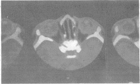
region revealed a picture of bilateral microophthalmia with retrolental soft tissue densities consistent with persistent hyperplastic primary vitreous syndromes (Fig. 1). The scan also revealed a
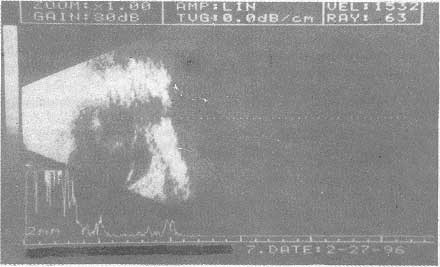
significant brain parenchymal atrophy. The picture of B-scan of the eye was also consistent with persistent primary hyperplastic vitreous
and exhibited bialteral retinal detachment (Fig. 2). The prognosis, depending on the above reports was explained and vitreous surgery was indicated. Detailed auditory examination was done which revealed sensori-neural deafness. The child was treated with phenobarbitone and intra- muscular magnesium, which decreased the frequency of flickerning movements. The child was. discharged.
 |
|
Fig. 1. CT
Scan
of cranio orbital region denoting bilateral micro-ophthalmia, persistent hyperplastic primary vitreous and brain parenchymal atrophy. |
Discussion
Norrie, a Danish Ophthalmologist surveying the causes of blindness in Denmark in 1927, described a particular type of congenital blindness as ' Atrophica oculi congenital'. Warburg, in 1961, proposed the name "Norrie's disease" for this condition of congenital bilateral pseudo-tumor of the retina with recessive X-linked transmission. The first case in India was described by Saini et al. in 1991(1). This is probably the second case reported.
Norrie's disease or Oculo acoustico cerebral degneration is usually inherited as a X-linked recessive disease with gene mapped on the short arm of chromosome X at the locus 11.3(2). All the affected patients are blind since birth. Mental subnormality occurs in about one third of the cases and 25-30% develop a sensori neural deafness. This usually presents with
leukocoria and deafness.
Variants of this disease can present with seizures as was seen in this case report. Apneic spells and nystagmus
are other presenting features. It could be associated with persistent hyperplastic primary vitreous, corneal opacities; cataracts and degenerative changes with phthisis bulbi.
Low levels of Mono-amine-oxidase A
and B are said to be the major factor in the pathogenesis of the disease(3). Mutations in the gene causing this disease have also been described. This could lead to a varied
presentation of the disease. A case with typical ocular features of Norrie's disease but. without mental retardation or hearing impairment has been described in Japan(4). Warburg suggested that the degenerative changes in the cerebrum and in the acoustic nerves are responsible for mental retardation and deafness.
 |
|
Fig. 2. B scan of the eye denoting persistent primary hyperplastic vitreous and retinal detachment. |
The histological picture of this disease is also characteristic(1,5). All the layers of vitreous are disorganized. Retinal dysplasia characterized by severe hypoplasia of the inner retinal layers and hyperplasia of retinal pigment epithelium with formation of rosettes is seen. Acoustic nerve degenerations as stated earlier, has now been found to affect the cochlear neurons and also the hair cells(5). B-scan of the orbit and CT scan help in confirming diagnosis as in this case and in assessing prognosis.
Early lensectomy, vitrectomy and retinal repair have been advocated for better
visual prognosis before total retinal detachment and phthisis. bulbi occurs. The family in this report refused all therapy.
An infant with leukocoria is an ophthalmic emergency. Congenital cataracts, retinoblastomas, retrolental fibroplasias, persistent hyperplastic primary vitreous and uveitis from Toxoplasmosis, Cytomegalovirus and Herpes form the differential diagnosis. Norrie's
disease, though a rare possibility should be considered in male infants with
bilateral retro-Iental masses.
This child presented with flickering of eyes, nystagmus and leukocoria, The detailed examination showed bilateral microophthalmia with bilateral persistent primary hyperplastic vitreous and retinal detachments and no response to either visual or auditory stimuli. These features clinically justified the diagnosis of Norrie's
disease which was confirmed by the CT scan and B-scan of the eye. The flickering
of the eyes were attributed to a low level of magnesium, which disappeared on
treatment with magnesium. This finding has not been reported earlier.
Familial incidences have been reported. Hence genetic counselling is advised in
affected families, along with antenatal ultrasound scan which can show retinal
detachment in affected cases around 20 weeks of gestation(6,7).
The aim of presenting this case is to highlight the features of Norrie's disease
and its variants. Affected families could be screened by DNA analysis and
appropriate action, like termination of pregnancy is carried out if detected antenatally, in the first trimester. This facility is currently not available in India. Genetic counselling is essential in all cases.
|
1.
Saini JS, Sharma A, Pillai P, Mohan K. Norries disease. Indian
J
Ophthal 1992; 40: 24-26.
2.
Sims KB, Lebo RV, Benson G, Shalish C, Schuback D, Chenzy Bruns G, et al. The Norrie's
disease gene Maps to 150 Kb region on chromosome Xp 11.3. Human Mol Genet 1992; 1: 83-89.
3.
Vossler DG, Wyler AR, Wilkus RJ, Gardner Walker G, Vicek BW. Cataplexy and Monoamine Oxidase deficiency in Norrie's disease. Neurology 1996; 46: 1258-1261.
4.
Isashiki X, Ohba-N, Yanagita T, Hokita N, Doi N, Nakagawa M, et al. Novel mutation at the initiation codon in the Norrie disease gene in two Japanese families Hum Genet 1995; 95: 105-108.
5.
Nadol JB Jr, Eavey RD, Liber Farb RM, Merchant SN, Williams R, Climenhager D, et al. Histopathology of the ears, eyes and brain in Norrie's disease. Oculo acoustico cerebral degeneration. Am
J
OtolaryngQl1990; 11: 112-124.
6.
Redmond RM, Graham CA, Kelly ED, Coleman-M, Nevin NC. Prenatal exclusion of Norrie's disease. Br
J
Ophthalmol 1992; 76: 491-493.
7.
Redmond RM, Vaughan JI, Jay M, Jay B. In utero diagnosis of Norrie's disease by ultrasonography. Opthalmic Pediatr Genet 1993; 14: 1-3.
|
