Pseudoaneurysm of any artery develops due to
collection of blood between its two outer layers, the tunica
media and the tunica adventitia. It is in contrast with the
true aneurysm which involves all three layers of the wall of
an artery. Among children sustaining traumatic injuries, 21%
have abdominal injuries [1,2]. Rarely, the blunt trauma of
the abdomen may be complicated by develop-ment of
pseudoaneurysm of hepatic artery, which may rupture inside
biliary tract, leading to life-threatening complication of
hemobilia. Classical signs of hemobilia consist of upper
abdominal pain, upper gastrointestinal hemorrhage and
jaundice, called Quincke triad. All these three signs are
present in only 22% of cases, whereas only upper
gastrointestinal bleeding is present in 42% of cases [3].
An 8-year-old child presented in our emergency
department with complaint of pain abdomen for 15 days and
hematemesis and melena for 10 days. The pain abdomen started
when he was punched in his abdomen by one of his
schoolmates. He took analgesics for his pain abdomen. There
was no history of fever, rash or any bleeding diathesis. He
was pale and had tachycardia at admission. There was no
history of fever, rashes or any bleeding diathesis. Blood
pressure was 113/70 mmHg and there was no petechial/purpuric
rash. He was given normal saline bolus and intravenous
pantoprazole followed by whole blood transfusion. Blood
investigations revealed low hemoglobin (4.8 g/100 mL) with
normal leucocyte counts, liver enzymes and renal function
tests; International normalized ratio was 0.95.
Ultrasonography abdomen done outside had revealed a 9 mm
calculus in gall bladder neck. Upper gastrointestinal
endoscopy, which had been done prior to coming to our
hospital, had documented erosion of mucosa of antrum and
pylorus with blood and blood clot inside stomach. Blood was
also seen coming out from ampulla of Vater and an impression
of erosive gastritis and hemobilia had been reported. The
child continued to have hematemesis after admission. A
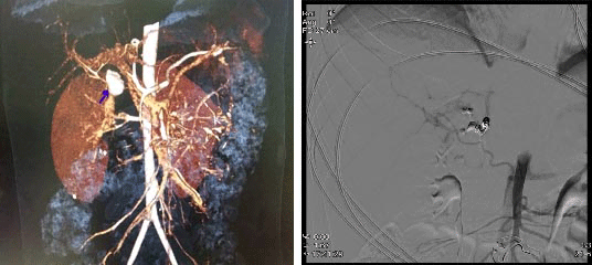
computed tomography (CT) angiography of the abdomen was done
which revealed a pseudoaneurysm of the right hepatic artery
(Fig. 1a). Percutaneous coil occlusion of
the right hepatic artery was done through the ipsilateral
femoral artery (Fig. 1b), and the hematemesis stopped
thereafter. He continued to have intermittent colicky pain
abdomen post procedure also, which persisted along with
melena, till sixth day of admission. The child became
completely asymptomatic on seventh day of admission, when he
was discharged. He was asymptomatic, without any pallor, and
with normal liver function test on follow up after one
month.
 |
| (a) |
(b) |
| Fig. 1
(a) Pseudoaneurysm of right hepatic artery in
CT-angiography (arrow), (b) Coil embolization of the
pseudoaneurysm of right hepatic artery. |
Approximately 1.7% of children sustaining blunt
trauma to the abdomen develop pseudoaneurysm of hepatic
artery and most of the pseudoaneurysm of the hepatic artery
are associated with the higher grades of liver injury [4].
Other causes of pseudoaneurysm of hepatic artery include
surgical procedures like cholecystectomy or percutaneous
procedures and endoscopic procedures like
cholangiopancreatography, liver biopsy and drainage of liver
abscess [5]. Pseudoaneurysm may produce mass symptoms and
local pain or the situation may be further complicated by
rupture of the pseudoaneurysm. Rupture of the pseudoaneurysm
occurs within days to weeks after the injury. When the
pseudoaneurysm ruptures inside the biliary system, it leads
to haemobilia and life threatening upper gastrointestinal
bleeding. Ultrasonography may demonstrate pseudoaneurysm as
a sac like structure with blood flow within it, but its
sensitivity is low (37%) although it has a high specificity
(100%). Contrast enhanced ultrasonography has been shown to
have high sensitivity (75%) and specificity (100%) [6].
Endoscopy may also detect hemobilia resulting from rupture
of pseudoaneurysm by demonstrating blood coming out from
papilla of vater, but it also carries a low sensitivity. CT
angiography is investigation of choice for pseudoaneurysm of
hepatic artery. It provides a precise location of the
pseudoaneurysm and delineates the involved blood vessel.
Percutaneous arterial embolization is highly effective
in controlling arterial bleeding in hemobilia [7]. Success
of endovascular management at experienced centres approaches
100% [8]. In a series of 176 children sustaining liver
injury, 3 (1.7%) had developed pseudoaneurysm of hepatic
artery [4]. Two of them experienced life-threatening
bleeding, both at 10 days after injury. This was controlled
by angiographic embolization in one and by laparotomy in
other. One asymptomatic patient underwent successful
embolization of a large pseudoaneurysm, seven days after
injury [4]. Hepatic necrosis, gall bladder ischemia, biliary
fistula and hepatic abscess are known complications of this
procedure. Surgical intervention is rarely necessary, and it
is usually reserved for failed percutaneous embolization.
However, it is first line of management if pseudoaneurysm is
infected or if it is compressing other vascular structures.
On follow-up of such children with coil embolization of
hepatic artery, clinical jaundice and liver function test
derangement should be looked for.
In conclusion, an
upper gastrointestinal bleeding associated with abdominal
trauma could be due to hemobilia due to ruptured
pseudoaneurysm of hepatic artery. It may lead to life
threatening hematemesis, hence prompt recognition of this
condition by CT angiography and its management is important.
Contributors: AP: drafted the manuscript, collected
clinical details; SK: was involved in doing percutaneous
coil occlusion of pseudoaneurysm of the patient in case
report; Abhiranjan P: did the literature search related to
the topic; PK: reviewed the article and suggested editing.
All authors reviewed article before final submission.
Funding: None; Competing interest: None stated.
REFERENCES1. Sharma M, Lakhoti
BK, Khandelwal G, Mathur RK, Sharma SS, et al.
Epidemiological trends of pediatric trauma: A single centre
study of 791 Patients. J Indian Assoc of Pediatr Surg.
2011;16:88-92.
2. Kundal V, Debnath P, Sen A.
Epidemiology of pediatric trauma and its pattern in urban
India: A tertiary care hospital-based experience. J Indian
Assoc Pediatr Surg. 2016;22:33.
3. Green MHA, Duell
RM, Johnson CD, Jamieson NV. Haemobilia. British J Surg.
2001;88:773-86.
4. Safavi A, Beaudry P, Jamieson D,
Murphy JJ. Traumatic pseudoaneurysms of the liver and spleen
in children: Is routine screening warranted? J Pediatr Surg.
2011;46:938-41.
5. Berry R, Han J, Kardashian Ani,
LaRusso NF, Tabibian JH. Hemobilia: etiology, diagnosis and
treatment. Liver Research. 2018;2:200-8.
6. Ren X,
Luo Y, Gao N, Niu H, Tang J. Common ultrasound and
contrast-enhanced ultrasonography in the diagnosis of
hepatic artery pseudoaneurysm after liver
transplan-tation. Exp Ther Med. 2016;12:1029-33.
7.
Saad WE, Davies MG, Darcy MD. Management of bleeding after
percutaneous transhepatic cholangiography or transhepatic
biliary drain placement. Tech Vasc Interv Radiol.
2008;11:60-71.
8. Fidelman N, Bloom AI, Kerlan RK Jr,
Laberge JM, Wilson MW, Ring EJ, et al. Hepatic arterial
injuries after percutaneous biliary interventions in the era
of laparoscopic surgery and liver transplantation:
Experience with 930 patients. Radiology. 2008;247:880-6.


