|
|
|
Indian Pediatr 2014;51: 335 |
 |
Lichen Scrofulosorum
|
|
Piyush Kumar, OP Jha and *Avijit Mondal
Departments of Dermatology, Katihar Medical College,
Bihar and *College of Medicine and JNM Hospital,
Kalyani, West Bengal, India.
Email: [email protected]
|
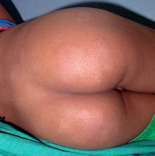
A 9-year-old boy presented with asymptomatic, skin colored,
grouped papules with mild scaling on elbows, buttocks,
knees, and dorsa of feet in a bilateral symmetric manner for
8 months (Fig. 1). There were no systemic
features and boy appeared otherwise healthy. Family history
was non-contributory. Mantoux text was positive (20 X 24
mm); routine blood investigations, chext X-ray and
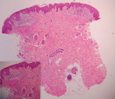
abdominal ultrasonography were normal. Histopathological
findings were consistent with lichen scrofulosorum (Fig.
2). He showed almost complete resolution of lesions
after four months of anti-tubercular therapy.
 |
|
Fig. 1 Symmetric skin
colored, grouped, papules with mild scaling on
buttocks.
|
 |
|
Fig. 2 Irregular acanthosis
and inflammatory cells in upper dermis (H&E X40);
Inset: non-caseating granuloma in upper dermis (H&E
X100).
|
Lichen scrofulosorum, an uncommon
tuberculid, is usually seen in children with nodal or
skeletal tuberculosis or following BCG vaccination. Lesions
are usually confined to the trunk and present as
asymptomatic, firm, follicular or perifollicular flat-topped
skin-colored or reddish brown papules, sometimes with fine
scales. Lesions may coalesce to form rough, discoid plaques,
and may persist for months. With anti-tubercular treatment,
the lesions usually clear within 12 weeks without scarring.
The differential diagnoses include lichen nitidus (more
shiny), lichen spinulosus, keratosis pilaris (keratotic
projections- antenna sign), phrynoderma, secondary syphilis,
papular sarcoidosis, pityriasis rubra pilaris, and
folliculitis. Histopathology is diagnostic and demonstrates
superficial granulomas around hair follicles and sweat
ducts, with little or no caseation necrosis.
|
|
|
 |
|
