Two children with VP shunt migration presented to us
interestingly almost at the same time, one week apart. The
first was a 14-month-old boy, who presented with fever,
cough and loose stool. He had undergone ventriculo
peritoneal (VP) shunt at the age of two months for
hydrocephalus following intraventricular hemorrhage . He was
a preterm baby with growth retardation and developmental
delay, receiving thyroxine and valproate. On examination, he
had open anterior fontanel with normal tension and patent VP
shunt. He had a reducible right indirect inguinal hernia and
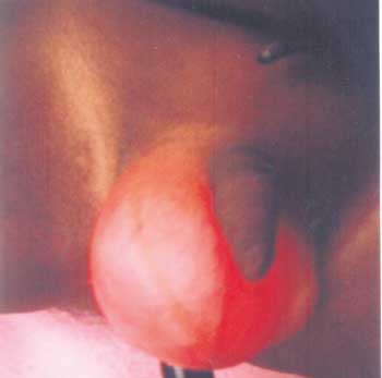
hydrocele. On the second day of admission, he developed
excessive intermittent crying and increase in size of
scrotal swelling. On transillumination, a cord like
structure was seen in the scrotum (Fig.1).
When the hernia was reduced, his crying stopped, but the
cord like structure persisted, which was clinically
diagnosed as VP shunt tube. CT head revealed moderate
hydrocephalus and VP shunt in situ. USG scrotum
showed hydrocele right side with VP shunt tube seen
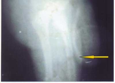
extending to the right scrotal sac. X-ray abdomen
showed shunt tube extending from abdomen into the scrotum (Fig.
2). Herniotomy and repositioning of VP shunt tube
were done.
 |
 |
|
Fig. 1 Transillumination of scrotum
showing VP shunt tube.
|
Fig. 2 X- Ray showing
migration of VP shunt into the scrotum.
|
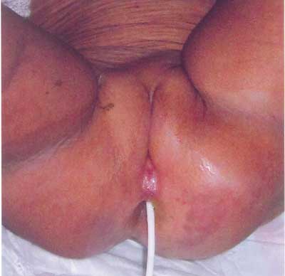
The second was a one-year-old female
child, with colpocephaly and partial corpus callosal
dysgenesis with hydrocephalus and VP shunt done 3 months
back. Subsequently, the child was treated for post-shunt
meningitis and peritonitis. She presented with abdominal
pain and extrusion of a tube like structure form the anal
orifice after passing stool, which was clinically diagnosed
as migrated VP shunt tube (Fig.3). She had
wide open, full anterior fontanel and patent VP shunt.
Laparotomy was done to reposition the tube and the distal
end was cut and extruded out per rectally.
 |
|
Fig. 3 Extrusion of VP shunt through the
rectum.
|
Migration of VP shunt is a known
complication, but extrusion into the genitalia and rectum
are rare.


