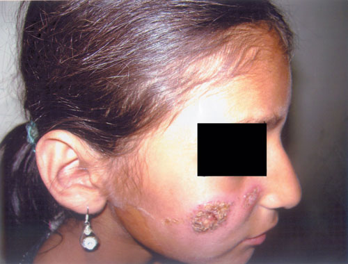
A 7-year-old girl presented for treatment of swelling over right cheek of
two months duration. On examination, she had several small, painless,
plaque lesions with indurated, erythematous and irregular borders and
central ulcerations evident on the right cheek (Fig 1).
There was no neurological deficit, or lymphadenopathy in the head and
neck. The medical history was not significant. Local biopsy on light
microscopy showed skin with hyperkeratosis, parakeratosis and acanthosis.
The dermis was filled with aggregates of large, pink, histiocytes, and
mixed chronic inflammatory cells. The histiocytes contained dot-like
organisms typical of LD bodies. She was treated with intramuscular sodium
stibogluconate for three weeks. The lesions disappeared a month later and
there has been no recurrence till the last follow-up.
 |
|
Fig.1 Plaque lesions with indurated and
irregular borders and central ulcerations. |
Differential diagnosis of localized cutaneous
leishmaniasis may include bacterial or fungal infections like impetigo,
lupus vulgaris, sporotrichosis or eczema. A chronic painless ulcer,
without any systemic symptoms in a child who has visited endemic region,
and not responding to routine treatment should suggest possibility of
cutaneous leishmaniasis.


