|
|
|
Indian Pediatr 2011;48: 321-323 |
 |
Umbilical Myiasis in Newborn |
|
Taraknath Ghosh, Kaustav Nayek, Nilanjan Ghosh and *Mrinal
Kanti Ghosh
From the Departments of Pediatrics and *Radiology,
Burdwan Medical College & Hospital, Burdwan, West Bengal, India.
Correspondence to: Dr Taraknath Ghosh, Doctors Qtr No 22,
block-3, Baburbag, Burdwan Medical College,
Burdwan, WB 713 104.
Email: [email protected]
Received: October 5, 2009;
Initial review: November 10, 2009;
Accepted: January 4, 2010.
|
Umbilical myiasis is rare in newborns. We are reporting two cases of
umbilical myiasis from rural West Bengal (India) that were infected by
larval forms of blow fly (Chrysomya megacephala). One of them
subsequently developed septicemia while the other one was clinically
well.
Keywords: Myiasis, Neonate, Umbilical.
|
|
M
yiasis
is an animal or human disease caused by the immature stage (maggots)
of flies which feed on the host’s necrotic or living tissue[1].
Myiasis may affect humans reared in poor hygienic conditions. It is
more common in children less than five years of age and with a rural
background [2]. Myiasis in the human neonatal period is a rare
occurrence and almost exclusively found in neotropic areas [3].
Case Report
Case 1: A nine-day-old, tribal male baby,
born at home out of non consanguineous marriage belonging to a poor
socioeconomic status from rural Bengal was admitted with complaints
of refusal to suck, and discharge from umbilicus. The baby was born
by normal vaginal delivery conducted by a local Dai, cried
immediately after birth. Antenatal period was uneventful. Mother had
received two doses of tetanus toxoid during pregnancy.
The baby weighed 2.1 kg and was lethargic, with
subnormal cry and reflexes. Temperature was slightly elevated. Vitals
were stable and anterior fontanel was soft and pulsatile. Abdomen was
slightly distended, liver and spleen was just palpable and soft. Foul
smelling purulent discharge from umbilicus with periumbilical flaring
was noted. On close observation after removal of pus with sterile
cotton swab, the tip of some white spindle shaped mobile worm like
structures were noted. These on pulling out with forceps proved to be
a maggot. Hemogram, urine and CSF analysis were normal. Ultrasound
scan of the umbilical area showed another twelve maggots and, as soon
as all larvae were out of the epidermis, the cellulites rapidly
resolved with no sequelae. Blood culture and culture from the
umbilical swab revealed growth of Staphylococcus aureus. The
baby was treated with intravenous cefotaxime and netilmycin for 7
days.
Case 2: A six day old female
neonate presented with history of something coming out from the
umbilical region. This baby was delivered vaginally at our medical
college. The intra and post natal events of mother were uneventful
and both mother and child were discharged from hospital on next day.
 |
|
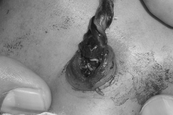
Fig. 1 A few maggots and partially
attached gangrenous cord at umbilicus of a newborn. |
On examination this neonate weighed 2.8 Kg. Her
cry, reflex and activities were satisfactory and all vital parameters
were within normal limit. The gangrenous cord was partially attached
at umbilicus and movements of some maggots were noted at the
umbilicus (Fig 1). In the next 3 hours, 13 maggots
bored out of umbilicus following instillation of ether (repellent).
Ultrasound examination showed another 6 maggots and they also were
removed similarly. Following removal the maggots were preserved in
80% of alcohol and sent to School of Tropical Medicine, Kolkata for
microscopic examination and species identification. The maggot was
found to be of Chrysomya megacephala. Blood samples were
collected from the neonate and all the reports were within normal
limits. There was no evidence of sepsis and the infant was discharged
from the hospital on the 3rd day under satisfactory physical and
clinical condition.
Discussion
Umbilical myiasis, a type of cutaneous tissue
myiasis, is usually produced by larvae of flies which are found in
wounds and gangrenous tissue where they act as facultative parasites
feeding on necrotic tissue and occasionally healthy tissue. In
umbilical myiasis the fly lays eggs on dry skin and the larvae
subsequently invade the wound and feed rapaciously on healthy tissue,
usually in groups to produce characteristic pocket like injuries.
Larvae grow rapidly and reach maturity in 4-8 days [4].
The larvae are removed from the affected site of
the host by irrigation, manipulation or surgery [5].
The larvae should be killed in hot water to retain the overall shape
as the posterior spiracles are very important for species
identification. Identification of the maggot can be crucial in
determining pathogenesis and as well as controlling of the disease.
Third stage larva is ideal for species identification [5].
Chrysomya megacephala, more commonly known as
the Oriental Latrine Fly, is known to breed in human feces, meat and
fish. In the rural Indian population, defecating in open air is a
common practice. The fly is attracted by feces and lays eggs on them.
After landing on feces it lands commonly on human foods and on very
rare occasion on open human wounds or on umbilicus of a newborn [6].
These may be the events that lead to umbilical myiasis in the two
cases that we have reported.
Acknowledgment: Dr AK Dutta, Associate
Professor, Pediatrics, Burdwan Medical College, for his participation
in the management of the cases, and Dr Netai Pramank, Assistant
Professor, School of Tropical Medicine, Kolkata, for expert comments.
Contributors: All were responsible for patient
care and investigation. TG & NG prepared the manuscript. KN organized
the management and follow up of the case. MKG was responsible for
radiological examination of the babies. All authors read and approved
the final manuscript.
Funding: None.
Competing interests: None stated.
References
1. Zumpt F. Myiasis in man and animals in the Old
World. 1st edition. London: Butterworths; 1965.
2. Singh I, Gathwala G, Yadav SP, Wig U, Jakhar KK.
Myiasis in children: The Indian perspective. Int J Pediatr
Otorhinolaryngol. 1993;25:127-31.
3. Duro EA, Mariluis JC, Mulieri
PR. Umbilical myiasis in a human newborn. J Perinatol.
2007;27:250-1.
4. Piangjai S, Siriwattanarungsee S,
Sukontason KL, Sukontason K. Morphology and developmental rate of
blowflies Chrysomya megacephala and Chrysomya rufifacies
in Thailand: application in forensic entomology. Parasitol Res.
2008;102:1207-16.
5. Cook GC, Zumla A. Medical Acarology and
Entomology. Manson’s Tropical Disease, 21st edition, Saunders (ELST);
2003. p. 1727-32.
6. Hammack L. Oviposition by screw-worm flies (Diptera:
Calliphoridae) on contact with host fluids. J Econ Entomol.
1991;84:185-90.
|
|
|
 |
|
