A 6-year-old boy, immunized for his age, presented with fever, malaise and
asymptomatic orocutaneous lesions for the past 3-4 days, after being
involved in cleaning activity of a fish pond. On examination he was
febrile (temperature 100ºF) with no lymphadenopathy. Oral cavity showed
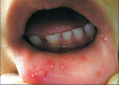
aphthae-like lesions having small yellowish-white shallow necrotic
vesicles surrounded by red areola over palate, buccal and labial mucosae
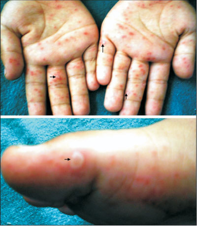
(Fig.1). Cutaneous lesions comprised multiple erythematous,
3-5mm, round/oval macules, and pearly-white vesicles with a red areola
over palms and soles (Fig.2), dorsal aspect of fingers,
buttocks and elbows. Systemic examination and investigative profile were
essentially normal. A clinical diagnosis of hand, foot and mouth disease
was made. The child was treated symptomatically.
 |
|
Fig.1 Small yellowish-white aphthae-like
lesions with surrounding erythematous areola seen over labial
mucosa. Similar lesions were present over buccal mucosa and anterior
palate. |
Hand, foot and mouth disease (HFMD) is a viral
infection usually caused by enteroviral genuses (usually coxsackie virus
A16, A5, A10, and sometimes coxsackie virus-B or human enterovirus 71).
HFMD is contagious usually spread via oro-fecal or respiratory routes. It
is a disease primarily affecting children, although the disease occurs
occasionally in adults. The diagnosis is primarily clinical by the
characteristic distribution of cutaneous lesions over hands, feet and
buttocks along with oral lesions. The cutaneous lesions begin as 3-7mm
erythematous macules evolving rapidly into pale white, oval, thin-walled
vesicles with a red areola. The lesions are typically elliptical, their
long axis parallel to the skin lines (Fig.2). They fade over
2-3 days and heal without crusting or scarring in about a week. Most HFMD
patients need only symptomatic treatment and reassurance in view of its
self-limiting benign clinical course.
 |
|
Fig.2 Small multiple erythematous,
round/oval macules and pearly-white vesicles with a red areola
(arrows) over palms and soles. |
In the absence of cutaneous lesions the oral lesions of
HFMD may be mistaken for aphthous ulcers, Herpes simplex
gingivostomatitis or oral varicella lesions. However, the oral erosions in
HFMD are usually smaller, more uniform and asymptomatic unlike those in
herpetic gingivosto-matitis which are painful and coalesces, and those of
varicella usually last longer and always crust. Unlike HFMD, both
varicella and herpes lesions will also show multinucleated giant cells in
Tzanck smears. Herpangina, another self limiting disease in children due
to multiple types of coxsackie viruses and echoviruses and characterized
by acute febrile illness with headache, sore throat, dysphagia, anorexia,
occasionally stiff neck, and small yellowish-white vesicles/ulcers with
erythematous areola distributed irregularly over posterior oropharynx
(anterior faucial pillars, tonsils, uvula, or soft palate), closely mimics
HFMD. However, absence of skin lesions and characteristic distribution of
oral lesions in herpangina are diagnostic. The skin lesions of HFMD can be
distinguished from Herpes simplex associated erythema
multiforme by the skin lesions which are round/oval, grey and targetoid.


