|
|
|
Indian Pediatr 2009;46: 354-356 |
 |
Juvenile Pagetís Disease |
|
CK Indumathi, Chitra Dinakar and Rakesh Roshan
From the Department of Pediatrics, St Johnís Medical
College Hospital, Bangalore, India.
Correspondence to: Dr CK lndumathi, Assistant Professoor,
Department of Pediatrics, St Johnís Medical College Hospital, Sarjapur
Road, Bangalore 560 034, India.
E-mail: [email protected]
Manuscript received: December 1, 2007;
Initial review completed: April 25, 2007,
Revision accepted: April 28, 2008.
|
|
Abstract
Juvenile Pagetís disease (JPD), a rare genetic
disorder characterized by markedly accelerated bone turnover, presents
in early childhood. We report a child with typical features of JPD who
remained undiagnosed till 15 years of age. Rarity of this disease in
Indian literature and need for early diagnosis to prevent progression of
disease prompted us to report this case.
Key words: Bisphosphonates Juvenile Pagetís disease, Metabolic
bone disease.
|
|
J
uvenile Pagetís disease (JPD) is an
autosomal recessive disorder characterized by increased bone turnover
secondary to enhanced osteoclastic activity(1). There are very few Indian
reports of JPD(2,3). We describe a 15-year old boy who presented with
characteristic features of JPD.
Case Report
A 15Ėyear old boy presented with the history of
progressive increase in head size and bowing deformity of the legs since
the age of 5 years. Problems started with a fracture and deformity of
right femur, which persisted despite surgical intervention. Subsequently,
he developed progressive diminution of vision in both the eyes. There was
no history of headache, vomiting or convulsions. He was a second child
born by normal delivery to second degree consanguineously wed couple.
Development was normal and he was attending school until he developed
diminution of vision. There was a family history of similar deformity of
limbs in his elder brother, which started around 6 years of age. At the
time of presentation, the elder sibling was 18 year old and immobile.
On examination, he weighed 55 kg. His height was 142 cm
(<3rd percentile) with upper segment, lower segment ratio of 0.76:1. He
had massive macrocephaly with a head circumference of 70 cm. Head was
asymmetrical. There was parietal pro-minence, depressed nasal bridge,
hypertelorism and left maxillary prominence. Teeth were malaligned. Hands
were broad and thick. There was severe genu valgum deformity and he could
walk only with support. Chest was broad and asymmetrical with prominent
thick clavicles and sternal prominence. There was no evidence of rickets.
Spinal examination revealed scoliosis to right. Respiratory and
cardiovascular systems were unremarkable. Per abdomen examination did not
reveal organomegaly. Neurological examination revealed normal intelligence
and normal motor examination. There was horizontal nystagmus with
bilateral optic atrophy and vision was limited to finger counting at 3
meters in both the eyes. Hearing was normal.
Investigations were as follows; Hb10g/dL, calcium 8.6
mg/dL and phosphorous 4.2 mg/dL. Alkaline phosphates was increased to 1799
units (normal=50-140 units).
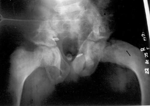
X-ray of pelvic bones and femur revealed lytic
lesions (Fig.1) with fracture and bowing. There was no
radiological evidence of rickets. X-ray skull revealed extensive sclerotic
lesions in the frontal bone and base of skull (Fig.2). CT
skull revealed thickening of medial wall of the orbit with optic canal
narrowing.
 |
|
Fig.1 X-ray pelvis revealing lytic lesions in the right femur
with extensive bowing.
|
 |
|
Fig. 2 X-ray skull lateral view revealing
extensive sclerotic lesions in the frontal bone and base of skull
anteriorly. There is evidence of bone enlargement with widening of
diploe. |
Diagnosis of JPD was made and the child was started on
oral alendronate 20 mg per day and calcitonin nasal spray 100 IU per day.
Decompression of the optic canal was considered. However, the procedure
was deferred in view of doubtful restoration of vision and anticipated
severe bleeding. At first follow-up after 4 weeks, alkaline phosphatase
level had started showing declining trend. Subsequently, he was lost to
follow-up.
Discussion
Pagetís disease is a metabolic bone disease
characterized by increased bone resorbing function of the osteoclast,
owing to an increase in its number, size and resorbing activity(1).
Initially it starts as "lytic phase" which is followed by compensatory
increased activity of osteoblasts "Sclerotic phase". The bone turnover
rate increases up to 20 times of normal. This results in increased bone
formation, which is more deficient, vascular and disorganized. As the new
skeleton is structurally weak and more vascular, patients present with
repeated fractures and deformities. Markedly elevated level of alkaline
phosphatase and radiographs revealing characteristic mosaic pattern (both
osteolysis and osteosclerosis) are diagnostic of Pagetís disease.
Pagetís disease is essentially a disease of adults
presenting in fourth and fifth decade of life(2). There are scarce reports
of JPD from India(3,4). Only 2 children below 16 years reported in a
series of 51 cases(3). JPD is characterized by widespread skeletal
involvement presenting in childhood with progressive deformities, short
stature, growth retardation, progressive macrocephaly and facial
deformity, mainly maxillary expansion. Index child had all these
characteristic features. Compression and trapping of nerves, especially
auditory and optic nerves result in deafness and optic atrophy. Our child
had optic atrophy but not deafness. Two other conditions considered in
differential diagnosis were polyostotic fibrous dysplasia and hereditary
hyperphosphatasia. Markedly elevated alkaline phosphatase level is unusual
in polyostotic fibrous dysplasia. Though, hereditary hyperphosphatasia is
associated with high level of alkaline phosphatase, it manifests much
earlier and radiographs do not reveal typical mosaic pattern.
Morbidity of JPD is very severe and majority of
children become wheel chair bound by 15 years if untreated(1,5). Oral
bisphosphonate therapy, especially alendronate, markedly suppresses bone
turnover and brings about remission(6-9). Early use in pediatric patients
prevents development of deformities and arrests progression of the disease
and further fractures. Though there is concern about permanent disturbance
of bone remodeling with alendronate, safe use has been documented in
pediatric age group in recent studies, at least in short term(10).
Calcitonin is a useful adjuvant, especially for pain relief(2).
Contributors: CKI was involved in the diagnosis and
management of the child and preparation of the manuscript. CD was involved
in the diagnosis and management of the child. RR was involved in
management of the case and preparation of the manuscript.
Funding: None.
Competing interest: None stated.
References
1. Deftos LJ. Treatment of Pagetís disease - taming the
wild osteoclast. NEJM 2005; 353: 872-875.
2. Joshi SR, Ambbore S, Butala N, Patvardhan M,
Kulkarni M, Pai B, et al. Pagetí disease from Western India. J
Assoc Physicians India 2006; 54: 535-538.
3. Anjalji Thomas N, Rajaratnam S, Shanthly N, Oommen
R, Seshadri MS. Pagetís disease of bone: Experience from a center in
Southern India. J Assoc Physicians India 2006; 54: 525-529.
4. Bhadada S, Bhansali A, Unnikrishnan AG, Khadgawat R,
Singh SK, Mithal A, et al. Does Pagetís disease exist in India? A
series of 21 patients. J Assoc Physicians India 2006; 54: 530-534.
5. Cundy T, Davidson J, Rutland MD, Stewart C, DePaoli
AM. Recombinant osteoprotegerin for Juvenile Pagetís disease. NEJM 2005;
353: 918-923.
6. Cundy T, Wbeadon L, King A. Treatment of idiopathic
hyperpbospbatasja with intensive bisphophonate therapy. J Bone Miner Res
2004; 19: 703-711.
7. Lombardi A. Treatment of Pagetís disease of bone
with alendronate. Bone 1999; 24 (5 Suppl): 598-618.
8. Wendlova J, Gabavy S, Paukovic J. Pagetís disease of
bone - treatment with alendronare, calcium and calcitriol. Vintr Lek 1999;
45: 602-605.
9. Khan SA, Vasikarnn S, McCloskey EV, Beneton MN,
Rogers S, Coulton L, et al. Alendronate in the treatment of Pagetís
disease of bone. Bone 1997; 20: 263-271.
10. Bianchi ML, Cimaz R, Bardare M, Zulian F, Lepore L, Boncompaign A,
et al. Efficacy and safety of alendronate for the treatment of
osteoporosis in diffuse connective tissue diseases in children. Arthritis
Rheum 2000; 43: 1960-1966. |
|
|
 |
|

