A 20-day-old female child born at term to a primigravida by normal
vaginal hospital delivery was admitted in neonatal intensive care unit
with respiratory distress. Immediate postnatal period was uneventful.
The baby had severe tachypnea, tachycardia and chest retractions. These
was no cyanosis and the heart sounds were audible on the left side of
the chest with grade II systolic murmur in second left parasternal area.
There was no evidence of any external congenital anomaly. Investigations
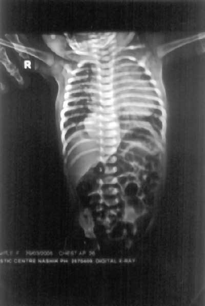
revealed normal hemogram, X-ray chest showed density in the right
paraspinal region along with medial inferior lung suggestive of
sequestration of right lower lobe (Fig. 1), no
cardiomegaly or dextroposed heart. Colour Doppler echocardiography
revealed ostium secundum ASD with anomalous pulmonary venous drainage.
CT scan of chest and abdomen with angiography showed complex cardiac
anomaly with aorta draining into the right lung. The consolidated right
lower lobe represented an area of sequestration. The patient underwent
coil repair of collateral to relieve pulmonary hypertension with
clinical improvement of symptoms.
 |
|
Fig. 1. Sequestration of right lower lobe.
|
The Scimitar syndrome is a rare condition of
cardiopulmonary anomalies(1) accounting for 0.5-1% of congenital heart
disease. The incidence of the associated congenital cardiovascular
abnormalities is 36% in pediatric age group and is highest (75%) among
the neonates and include ASD, VSD, coarctation of aortic arch and
abnormal relationship of the pulmonary arteries and bronchi(2). The age
of presentation is variable. In a series of 32 patients over a period of
20 years, the median age at diagnosis was 7 months(3). Severe
respiratory insufficiency is always present in symptomatic cases in
early age group as Scimitar syndrome results in pulmonary hypertension,
heart failure and right lung infection. In older children and adults,
the diagnosis of Scimitar syndrome is often made incidentally who
undergo chest radiography for diverse reasons. Recurrent respiratory
infections and heart murmur may be the mode of presentation in them.
Children who are diagnosed with Scimitar syndrome after infancy have
fewer associated defects and less pulmonary hypertension.
Treatment for symptomatic Scimatar syndrome consists
of surgical repair. Repair of the anomalous venous return and ligation
of colloaterals is generally recommended, although right pneumonectomy
also provides similar results(4).
Our patient responded for 6 weeks but then
deteriorated and therefore was referred to cardio-thoracic center in
Mumbai for further surgical management.
S.S. Kashyape,
A.M. Tilak,
Shree Medical Research Center,
c/o. Kashyape Children Hospital,
Opp. Hotel Mazda, Trimbak Naka,
Nasik 422 002, Maharashtra,
India.
1. Canter CE, Martin TC, Spray TL, Weldon CS,
Strauss AW. Scimitar syndrome in childhood. Am J Cardiol 1986; 58:
652.
2. Gilonyo DK, Tandon R, Lucas RV Jr, Edwards JE.
Scimitar syndrome in neonates: report of four cases and review of the
literature. Pediatr Cardiol 1986; 6: 193-197.
3. Najm HK, Williams WG, Coles JG, Rebeyka IM.
Freedom RM. Scimitar syndrome: twenty year’s experience and results of
repair. J Thorac Cardiovasc Surg 1996; 112: 1161-1168.
4. Huddleston CB, Exil V, Canter CE, Mendeloff En. Scimitar
syndrome presenting in infancy. Ann Thorac Surg 1999; 67: 154-159.
