|
|
Images in Clinical Practice Indian Pediatrics 2006; 43:349-350 |
||||
|
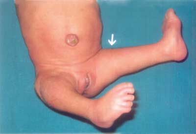
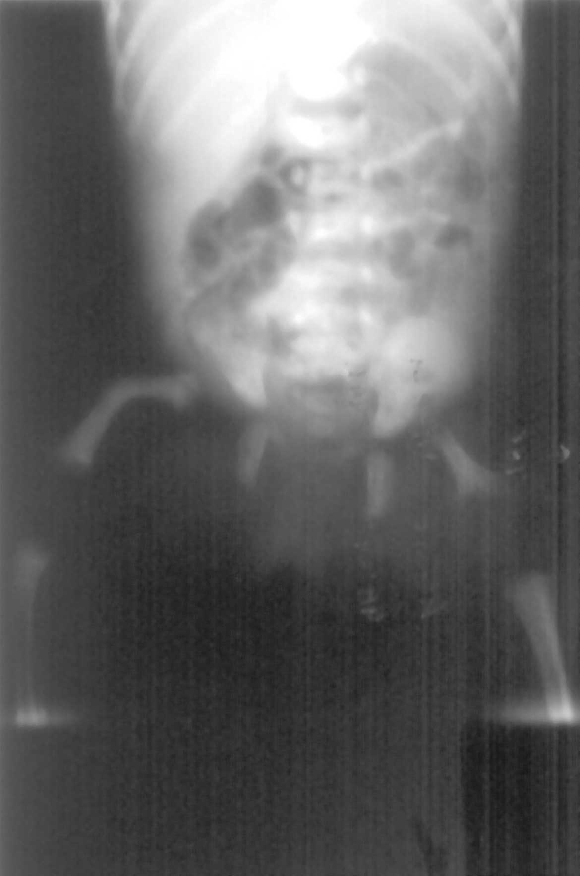
Proximal Femoral Focal Deficiency |
||||
|
Proximal femoral focal deficiency (PFFD) is a rare anomaly with an estimated incidence of <0.2/10,000 births, with bilateral deficiency in 10 to 15% of cases. Nutritional disturbance or vascular insult to the mesenchymal tissue during femoral embryogenesis is postulated as a possible etiology. No known inheritance but recurrence in sibs have been reported. PFFD is almost always an isolated occurrence except for associated ipsilateral fibular hemimelia. It varies in severity from a marginally short femur to a complete absence of femur in severe cases. Kyphomelic dysplasia (rhizomesomelic shortening with lateral bowing of the long bones, flared irregular metaphyses, a short narrow chest, mild dysmorphic facial features) and campomelic dysplasia (macrocephaly, hypertelorism, long philtrum, micrognathia, hypotonia, hypoplastic scapulae, bowing of the long bones, vertical narrow iliac bones, and absence of ossification of the thoracic pedicles) are important differential diagnosis. Prognosis depends on the degree of hypoplasia. Periodic accurate clinical and radiological examinations are necessary to document the character of the deformities, growth and bone development of femur, and function of the extremity. This will determine the need for surgery and prosthetic supplementation. Prenatal sonography is useful both for detecting PFFD and separating it from syndromes showing global skeletal abnormalities. Ankit Parakh,
|
![]()