|
|
Recommendations Indian Pediatrics 2001; 38: 975-986 |
||||||||||||||||||||||||||||||||||||||||||||||||||||||||||||||||||||||||||||
|
Consensus Statement on Management of Steroid Sensitive Nephrotic Syndrome |
||||||||||||||||||||||||||||||||||||||||||||||||||||||||||||||||||||||||||||
Nephrotic syndrome is an important chronic disorder in children. About 90% children with idiopathic nephrotic syndrome have ‘minimal lesion’ on renal histological examination and respond promptly to corticosteroid therapy with remission of proteinuria. Approximately three-fourths of these patients have one or more relapses that require repeated treatment with cortico-steroids. Such patients are at high risk of corticosteroid toxicity, frequent serious infections and other complications. A small proportion of patients who are steroid resis-tant are also at risk for similar complications and renal insufficiency. Most pediatricians would encounter only a few children with nephrotic syndrome in their practices. In view of the complexity of treat-ment, early referral to a pediatric nephro-logist is recommended. Thereafter, long-term management of a child with nephrotic syndrome should be a collaborative effort between the primary pediatrician and pediatric nephrologist. An Expert Group Meeting of the Indian Pediatric Nephrology Group was held on 15 December 2000 at New Delhi to formulate recommendations for the management of patients with nephrotic syndrome (Annexure 1). This statement refers to the diagnosis, initial evaluation and management of steroid responsive nephrotic syndrome. Definitions Nephrotic syndrome is characterized by heavy proteinuria, hypoalbuminemia (serum albumin <2.5 g/dl), hyperlipidemia (serum cholesterol >200 mg/dl) and edema(1). Proteinuria is considered to be in the nephro-tic range when the urine protein is 3+/4+ on a dipstick test, spot protein/creatinine ratio >2 mg/mg, or urine albumin >40 mg/m2 per h (on a timed sample). In most instances, the finding of 3+/4+ proteinuria (on dipstick or boiling test) is adequate for defining nephrotic range proteinuria. Precise quantitative assessment of proteinuria is not essential and a 24-h urine protein measurement is not required for the diagnosis of nephrotic syndrome. Definitions useful in defining the course of the disease are shown in Table I(1). Initial Evaluation The diagnosis of nephrotic syndrome is based on the presence of heavy proteinuria, hypoalbuminemia and edema. Once the diagnosis is made, a detailed evaluation of the patient is necessary before starting treatment with corticosteroids. The height, weight and blood pressure are recorded. A regular weight record, during a relapse, helps monitor the decrease of edema until dry weight is achieved. Physical examination to detect infections is necessary, which if present should be treated promptly. Patients should be examined for an underlying systemic dis-order, e.g., systemic lupus erythematosus, amyloidosis and Henoch Schonlein purpura. Screening investigations to be carried out at the initial episode are: (a) Urinalysis; (b) Complete blood count, blood levels of albumin, cholesterol, urea and creatinine; (c) Blood levels of antistreptolysin O and C3 in patients with persistent microscopic or gross hematuria; (d) Appropriate tests for under-lying illness if clinically suspected (e.g., antinuclear antibodies for systemic lupus erythematosus); (e) Urine culture, if urinary tract infection is suspected; (f) X-ray chest, Mantoux test; and (g) Hepatitis B surface antigen. Table I - Definitions
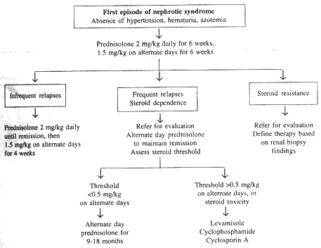
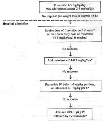
Treatment of Initial Episode Adequate treatment of the initial episode is extremely important. Current evidence suggests that treatment of the initial episode influence the subsequent course of the illness(2). The intensity of initial treat- ment may decrease the rate of subsequent relapses. It is necessary to treat infections before starting treatment with prednisolone. Medication Prednisolone is the standard medication for inducing remission. Other drugs such as dexamethasone, betamethasone and hydro-cortisone should not be used. Deflazocort, a steroid analogue, is as effective as predniso-lone and has fewer corticosteroid side effects but has not been widely employed (it is not available in India). Treatment Regimen Various treatment regimens have been used for the treatment of the initial episode of nephrotic syndrome. The International Study for Kidney Diseases in Children had origi-nally recommended four weeks each of daily and alternate day steroid therapy(3) which was almost universally used until recently. The available literature was considered by the Expert Group(1-7). The Expert Group recommends that the initial episode be treated with prednisolone administered in a dose of 2 mg/kg (maximum 60 mg) in two-three divided doses daily for six weeks, followed by 1.5 mg/kg (maximum 40 mg) as a single morning dose on alternate days for the next six weeks. Treatment with prednisolone is then discontinued. Cortico-steroids should preferably be administered after meals. Some workers recommend that alternate day treatment not be stopped abruptly, but gradually tapered over 6 months. A recent meta-analysis shows that extension of therapy to 3-6 months is associated with prolonged remission and reduced frequency of relapses(2). A longer regimen using 8 weeks daily and 8 weeks alternate day cortico-steroids was found useful but steroid toxicity was also higher(7). Other regimens are being evaluated at several centers and it is likely that a different regimen might emerge after a few years. Treatment of Relapse The patient should be examined for infections, which are treated before initiating corticosteroid therapy. Prednisolone is admi-nistered in a dose of 2 mg/kg/day (single or two divided doses) until urine protein is trace or nil for 3 consecutive days, or for two weeks. Subsequently, prednisolone is given in a dose of 1.5 mg/kg on alternate days for 4 weeks, and then discontinued(8). The usual duration of treatment for a relapse is thus 5-6 weeks. Prolongation of therapy is not necessary for patients with infrequent relapses (see below). In case the patient is not in remission despite two weeks treatment with daily pred-nisolone, such treatment might be extended for two more weeks. Patients requiring daily corticosteroid therapy for more than 2 weeks, to induce remission, should be referred to a pediatric nephrologist for evaluation. Infrequent Relapsers Patients who have three or less relapses a year and respond promptly to prednisolone are managed using the aforementioned regimen for each relapse. Such children are at a low risk for developing steroid toxicity. Frequent Relapsers or Steroid Dependence Patients with frequent relapses or steroid dependence should be managed in consulta-tion with a pediatric nephrologist. It is not usually necessary to perform a renal biopsy in these cases. Following induction of remission, the dose of prednisolone is tapered gradually to maintain the patient in remission on alternate day dose of 0.5 mg/kg or lower. Alternate day prednisolone may be administered for 9-18 months. A close monitoring of growth and blood pressure, and evaluation for features of steroid toxicity is essential. If the pred-nisolone threshold, to maintain remission, is higher than 0.5 mg/kg on alternate days, the following immunomodulators are recommended. (a) Levamisole may be administered in a dose of 2 -2.5 mg/kg on alternate days for 12- 24 months(9). Treatment with predniso-lone, 1.5 mg/kg on alternate days, is continued. The dose of prednisolone is gradually reduced by 0.15-0.25 mg/kg every 4 weeks to a maintenance dose of 0.25 mg/kg, which may be continued for six months. The chief side effect of treatment with levamisole is leukopenia; flu-like symptoms and skin rash may occur rarely. The total leukocyte count should be monitored every 4-8 weeks. (b) Alkylating agents, cyclophosphamide (2 mg/kg/day) or chlorambucil (0.1-0.2 mg/kg/day), may be administered along with alternate day prednisolone (1-1.5 mg/kg) for 12 weeks. Only a single course of either of the medications is recommended. In view of larger experience and safer toxicity profile, cyclophosphamide is usually preferred(10). The total leukocyte count should be monitored every 2-3 weeks. Treatment is discontinued if the leukocyte count falls below 4000/mm3. Increasing the fluid intake and frequent voiding can prevent hemorrhagic cystitis. Treatment with cyclophosphamide may rarely cause alopecia, nausea and vomiting. (c) Cyclosporin is administered in a dose of 4-5 mg/kg daily for 12-24 months. Therapy is continued with prednisolone in a dose of 1.5 mg/kg on alternate days for 4 weeks, and than tapered gradually (see leva-misole, above). Side effects such as hypertension, gum hypertrophy and hirsutism may occur, in which case the dose of cyclosporin may be reduced to 3 mg/kg/day. Relapses during and following treatment with levamisole, cyclophosphamide and cyclosporin are treated with the standard regimen (treatment of relapse, above). The advantages of using these drugs should be balanced against their potential toxicity. Levamisole has a weak steroid sparing effect and is useful in milder cases. Treatment with cyclophosphamide may be considered in patients showing: (i) significant steroid toxicity, (ii) severe relapses with hypovolemia or thrombosis, and (iii) poor compliance or follow up. Cyclosporin is recommended for patients that continue to show steroid dependence or frequent relapses despite a course of levamisole and cyclo-phosphamide. In some patients receiving treatment with levamisole or cyclosporin, corticosteroid treatment may be discontinued after six months. A protocol summarizing the management of patients with steroid sensitive nephrotic syndrome is shown in Fig. 1. Supportive care This forms an important aspect of managing children with nephrotic syndrome. Diet A balanced diet adequate in protein and calories is recommended. The child should receive 1.5-2 g/kg of proteins. Patients with persistent proteinuria are prone to mal-nutrition and should receive 2-2.5 g/kg of protein daily(5). Not more than 30% calories should be derived from fat and saturated fats avoided. Carbohydrates are best given as complex forms (starch and maltodextrin). Salt restriction is not necessary in most patients with steroid responsive nephrotic syndrome. A modest reduction (1-2 g per day) is advised in the presence of marked edema. Salt should not be added to salads and fruits; snacks containing high salt are avoided during these periods. Corticosteroids stimulate the appetite, and advice should be given about ensuring physical activity and preventing excessive weight gain. Edema Control of edema is an integral part of supportive care. Treatment with cortico-steroids usually leads to diuresis within 48-72 hours. Diuretics are thus avoided unless edema is significant and should not be used in children with diarrhea, vomiting or hypo-volemia. In moderate or persistent edema, frusemide is administered in a dose of 1-3 mg/kg per day. Additional treatment with potas-sium sparing diuretics (e.g., spironolactone) is not required if frusemide is used in this dose for less than one week. Patients requiring higher doses and prolonged duration of treatment with frusemide should receive spironolactone (dose 2-4 mg/kg daily). Blood pressure should be monitored frequently. A gradual reduction of edema, over one week, is preferred(11). Edema not responding to the above therapy should be managed in a hospital under close supervision. For refractory edema, a combination of diuretics and albumin infusion may be used. Infusion of albumin is followed by administration of frusemide in a dose of 1-2 mg/kg intravenously. Though infusion of albumin results in increased urine output, the effect is not sustained, especially in patients with steroid resistant nephrotic syndrome. Albumin infusions are usually administered on alternate days to allow fluid shifts to occur and prevent fluid overload. Patients receiving albumin should be carefully observed for respiratory distress and congestive heart failure. Refractory ascites interfering with respiration or associated with breaks in the skin may be removed by repeated tapping. A protocol for treatment of edema is shown in Fig. 2.
Fig. 1. Management of patients with steroid sensitive nephrotic syndrome. Patient and Parent Education Adequate information about the disease, associated complications and the expected course should be provided. Parents should be reassured that despite a relapsing course, progression to end stage renal failure necessitating dialysis or transplantation is extremely rare. Parental motivation and involvement is essential in management of a child with nephrotic syndrome. The following measures are emphasized. (a) Urine examination for protein at home using a dipstick, sulfosalicylic acid or boiling test. The examination should be done every morning during a relapse,
Fig. 2. Management of edema in patients with nephrotic syndrome.
Other Medications Antacids or histamine receptor (H-2) antagonists e.g., ranitidine need not be administered routinely in patients on treatment with corticosteroids. Children with upper gastrointestinal discomfort may how-ever be given the steroid dose along with an antacid. Calcium supplementation is usually not necessary unless the patient is on long-term steroids. Patients with steroid responsive nephrotic syndrome do not require medica-tions for hyperlidemia, since blood lipid levels rapidly normalize following remission. Immunization Parents should be advised regarding the necessity of completing the primary immunization. Immunocompromised patients should not receive live attenuated vaccines; inactivated or killed vaccines are safe(12). As a general guideline, patients receiving pred-nisolone in a dose of 2 mg/kg per day or greater, or total 20 mg/day or greater (if weighing more than 10 kg) especially when given for more than 14 days are considered immunocompromised. Live vaccines should be administered once the child is off steroids for 6 weeks. These vaccines may, however be given to patients receiving alternate day prednisolone at a dose less than 0.5 mg/kg, if there is a pressing need. Children with nephrotic syndrome are susceptible to infections, with encapsulated organisms. Parents should be explained about the advantages of vaccines against Strepto-coccus pneumoniae, Haemophilus influenzae, varicella and hepatitis B. Pneumococcal vaccination is recommended in all children with nephrotic syndrome, especially those with a previous episode of peritonitis(13). The vaccine should be given during remission and preferably when the child is not receiving daily prednisolone. A booster dose is recom-mended every five years for those children who have had the initial pneumococcal vaccine before the age of five years but continue to relapse(13). Patients in remission and not on corticosteroid therapy should receive varicella vaccine in a two-dose schedule, administered four weeks apart. Kidney Biopsy Most children with nephrotic syndrome not having hematuria, hypertension or impaired renal function are treated initially without undergoing a kidney biopsy. It is necessary to identify the underlying renal disease in certain cases (Table II)(1). A biopsy is not usually indicated in patients with frequent relapses when treatment with levamisole or cyclophosphamide is being considered. However, a biopsy should be performed before starting therapy with cyclosporin. Table II__Indications for Kidney Biopsy
After initial treatment
An expert should perform the biopsy. Centers that perform kidney biopsies should have facilities for evaluation of the specimens by light and immunofluorescence microscopy. Referral to Pediatric Nephrologist A pediatrician can adequately manage most patients with steroid responsive infrequently relapsing nephrotic syndrome. In all other circumstances, it is advisable to treat the patient in collaboration with a pediatric nephrologist (Table III). Complications Pediatricians should be aware of the complications that may occur. These compli-cations may develop in steroid responsive as well as steroid resistant nephrotic syndrome. Early diagnosis helps in the timely and accurate management of these problems. Infections in Nephrotic Syndrome Children with nephrotic syndrome are susceptible to severe infections, which need prompt treatment. Common infections include peritonitis, cellulitis and pneumonia. Viral and bacterial infections may precipitate relapses. The clinical features and management of common serious infections are shown in Table IV. Varicella may be a severe illness in patients with nephrotic syndrome who are immunocompromized. Patients who are unimmunised and exposed to a case of chickenpox are recommended prophylaxis with varicella zoster immunoglobulin(14). This preparation is expensive and not easily available in India. Immunocompromized patients who develop varicella should be treated with oral or intravenous acyclovir depending on the severity of the infection. Treatment with steroids is discontinued. Patients with nephrotic syndrome, who are Mantoux positive but show no evidence of disease, should receive prophylaxis with INH and rifampicin for six months(15). Those having evidence of active tuberculosis should receive standard antitubercular therapy for two weeks before starting corticosteroid treatment for nephrotic syndrome. Antibiotic prophylaxis: Patient with steroid responsive nephrotic syndrome and massive ascites may receive prophylaxis with penicillin V in a dose of 125 to 250 mg twice daily until the ascites has resolved(16). However, there is no evidence that routine application of antibiotic prophylaxis reduces the occurrence of serious infections in these patients.
TABLE IV–Clinical Features and Management of Common Infections
* Initial therapy may be parenteral for 5 days; once patient is nontoxic and accepting orally,
the medication may be administered orally.
Thrombotic Complications Children with nephrotic syndrome are at risk for venous and, rarely, arterial throm-bosis(12). Reduced intravascular volume and other abnormalities predispose to thrombus formation. Diuretics should be used judi-ciously. Puncture of deep vessels should not be done. Renal vein thrombosis is suspected in a patient with oligoanuria, hematuria or flank pain especially following an episode of dehydration. Ultrasound examination of the abdomen might show large kidneys and thrombi in renal veins. Femoral arterial thrombosis may occasionally occur. Deep vein thrombosis of calf veins is less common in children but may lead to pulmonary embolism. Saggital sinus and cortical venous thrombosis may follow episodes of diarrhea and present with convulsions, vomiting, altered sensorium and neurological deficits. Doppler studies and cranial CT scan are useful in confirming the diagnosis. Patients with thrombotic complications require urgent treatment under the supervision of a specialist. The treatment is supportive and consists of early mobilization, treatment of sepsis, prompt treatment of dehydration and cautious use of anticoagulants(5). Hypertension This may be noted at the onset of nephrotic syndrome or occur due to steroid toxicity. Therapy may be initiated with ACE inhibitors, calcium channel or b adrenergic blockers. Hypovolemia This complication can occur due to unsupervised use of diuretics especially if accompanied by septicemia, diarrhea or vomiting. The diagnosis is suggested by the presence of hypotension, tachycardia, cold extremities and poor capillary refill; blood levels of urea and uric acid are elevated. Some children might complain of moderate to severe abdominal pain. A rapid infusion of normal saline or plasma in a dose of 15-20 ml/kg, or albumin 1 g/kg is essential. The blood pressure should be monitored carefully. Albumin should be used with caution if the child is hypertensive because of the risk of pulmonary edema. Once adequate hydration is achieved, but the child remains oliguric, a single dose of frusemide (1-2 mg/kg intravenously) may be given. In case no urine is passed despite these measures, the diagnosis of acute renal failure is suspected. Steroid Side Effects Prolonged high-dose corticosteroid therapy may be associated with significant side effects. Parents and the child (if old enough to understand) should be explained about the side effects of corticosteroids. Steroids result in an increased appetite, cushingoid features, impaired growth, behavioral changes, gastritis, salt and water retention, hypertension and bone deminerali-zation. Children on prolonged treatment with corticosteroids should be monitored for steroid toxicity. Examination for cushingoid features, monthly record of blood pressure, six-monthly record of height and weight, and yearly evaluation for cataract is recommended. Steroids during stress Patients who have received high-dose steroids for more than two weeks in the past one-year show suppression of the hypo-thalamo-pituitary-adrenal axis. These children require supplementation of steroids during surgery, anesthesia or serious infections(17). Treatment with corticosteroids should not be stopped in patients having serious infections. Corticosteroid supplementation during stress is usually in the form of parenteral hydro-cortisone, which is administered in a dose of 2-5 mg/kg/day, followed by prednisolone 0.5 -1 mg/kg/day once the child is able to accept orally. This dose is administered for the duration of stress then tapered daily by 50%. Steroid Resistant Nephrotic Syndrome Patients showing resistance to oral corticosteroids pose problems in manage-ment. They usually show an unsatisfactory response to levamisole and alkylating agents. Most develop refractory edema and hyper-tension. Complications including repeated infections and malnutrition are common. These patients are at risk for developing chronic renal failure. Such children should be managed in collaboration with a pediatric nephrologist. A kidney biopsy should be performed to determine the underlying pathology. Patients showing minimal change disease or focal segmental glomerulosclerosis might benefit from treatment with high- dose intravenous corticosteroids or cyclosporin(12). Patients with persistent proteinuria benefit from treatment with ACE inhibitors, which reduce the severity of proteinuria in most cases. Therapy may be started with enalapril in a dose of 0.2-0.4 mg/kg in two divided doses with a careful watch on blood levels of creatinine and potassium. Diuretics are administered to control massive edema. Measures should be taken to prevent infections, control hypertension and reduce low-density lipoprotein cholesterol. Blood level of urea, creatinine, electrolytes, albumin and cholesterol are monitored. Conclusions These recommendations represent the consensus view of the Indian Pediatric Nephrology Group. They have been formu-lated on basis of best current practice, which is supported by studies published in peer-reviewed journals and the experience of the Expert Group. They are intended to provide pediatricians with broad guidelines for managing children with nephrotic syndrome. Therapy needs to be individualized for each patient and optimal care will be achieved by the combined inputs of a pediatrician and a pediatric nephrologist. Annexure I Participants of the Expert Group Meeting
Compiled by: Arvind Bagga, M. Kanitkar, R.N. Srivastava on behalf of Indian Pediatric Nephrology Group. | ||||||||||||||||||||||||||||||||||||||||||||||||||||||||||||||||||||||||||||
| References | ||||||||||||||||||||||||||||||||||||||||||||||||||||||||||||||||||||||||||||
|
![]()