|
|
|
Indian Pediatr 2012;49: 760
|
 |
Panophthalmitis in Dengue Fever
|
|
Siva Saranappa SB and *HN Sowbhagya
Departments of Pediatrics and *Ophthalmology,
Kempegowda Institute of Medical Sciences Bangalore, Karnataka.
Email:
[email protected]
|
|
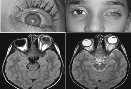
We report a rare case of panophthalmitis in dengue fever. A 6 year old
girl presented with 5 days fever and rash on the lower extremities,
flushed face and myalgia. Examination revealed fever of 38ºC,
erythematous rash, and flushed face. Investigations revealed leucopenia
and thrombocytopenia. Dengue serology was positive (both IgM and IgG).
Ultrasound showed ascitis and right pleural effusion. Next morning, she
complained of severe pain in the left eye. On examination, right eye was
normal. Vision in the left eye was 6/18. Fundus examination revealed
focal leaks of exudates from venous ends of retinal vessels at superior
quadrant. By evening, the left eye was swollen, with intolerable pain
and vomiting. Examination revealed shallow anterior chamber, intense
ciliary congestion, cloudy cornea; percluding evaluation of underlying
details. Vision was reduced to perception of light. Intraocular pressure
was increased. A diagnosis of acute angle-closure glaucoma was suspected
and was treated accordingly. By next morning, there was gross loss of
vision, proptosis and corneal clouding. Anterior chamber showed
organized disc of exudates. Panophthalmitis was suspected and ultrasound
was done which revealed thickening of choroid, sclera and exudation of
vitreous. MRI confirmed the diagnosis and showed diffuse inflammatory
thickening of the left ocular coats with hazy vitreous, peri-ocular
extensions of the inflammatory process involving both pre and post
septal soft tissues, retro-orbital fat and peri-optic neural sheath
showed inflammatory changes.
 |
|
Fig.1 Showing cloudy cornea, and MRI
sections of panophthalmitis.
|
Ocular manifestations in dengue, though rare, are not
uncommon, with 20% having ocular pain [1] and 40.3% having
subconjunctival hemorrhage, dilatation and tortuosity of retinal vessels
and hard exudates [2]. Chorioretinitis, retinitis, retinal vasculitis
and optic nerve involvement have been found to be associated with dengue
[3]. Anti-IgM dengue antibody was found to be positive in 18% of
patients with multifocal retinitis [4]. The triad of eye flashes,
floaters and blurring of vision was highly predictive for the
development of retinal hemorrhages [5]. The pathogenesis of
panophthalmitis is not known. It could be the part of immunologic and
inflammatory response to the dengue virus infection. The child
recovered, with visual loss of left eye.
Ocular manifestations in dengue are rare, but can be
as serious as panophthalmitis in this child. So a systematic ophthalmic
examination in patients with dengue fever, especially with ocular
symptoms, is mandatory.
References
1. Humayoun MA, Waseem T, Jawa AA, Hashmi MS, Akram
J. Multiple dengue serotypes and high frequency of dengue hemorrhagic
fever at two tertiary care hospitals in Lahore during the 2008 dengue
virus outbreak in Punjab, Pakistan. Int J Infect Dis. 2010;14:e54-9.
2. HK Kapoor, Saloni B, Mary J. Ocular manifestations
of dengue fever in an East Indian epidemic. Can J Ophthalmol.
2006;41:741-6.
3. Khairallah M, Chee SP, Rathinam SR, Attia S,
Nadella V. Novel infectious agents causing uveitis. Int Ophthalmol.
2010;30:465-83.
4. Shukla J, Saxena D, Rathinam S, Lalitha P, Joseph
CR, Sharma S, et al. Molecular detection and characterization of
West Nile virus associated with multifocal retinitis in patients from
southern India. Int J Infect Dis. 2012; 16:e53-9.
5. Seet RC, Quek Am, Lim EC. Symptoms and Risk Factors
of ocular complications, following Dengue Infection. J Clin Virol.
2007;38:101-5.
|
|
|
 |
|
