| |
|
Images Indian Pediatrics 2008; 45:783-784 |
||||||
|
Larsen Syndrome |
||||||
|
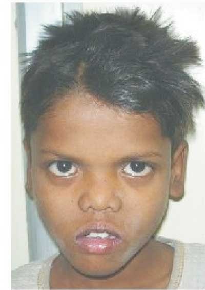
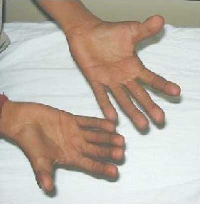
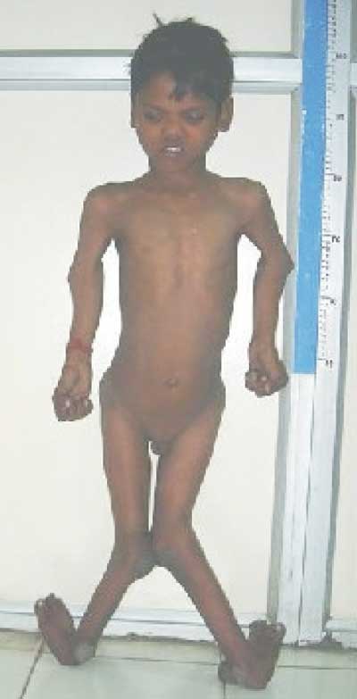
Larsen syndrome (OMIM 150250) is a complex syndrome with genetic heterogeneity, and with both autosomal dominant and autosomal recessive patterns of inheritance. Mutations in gene encoding filamin B (FLNB) result in Larsen syndrome. This gene has an important role in vertebral segmentation, joint formation and endochondral ossification and is also mutated in atelosteogenesis types I and III, and in spondylocarpotarsal syndromes. Autosomal dominant form is characterized by flat facies, joint hypermobility, congenital multiple joint dislocations, especially of the knees and talipes equinovarus. The mid-face is hypoplastic with a depressed nasal bridge. Cleft palate may be present. Osteoarthritis involving large joints and progressive kyphoscoliosis are potential complications. Airway obstruction caused by tracheomalacia and bronchomalacia may be life threatening. All affected individuals should be evaluated for cervical spine instability and caution should be taken during anesthesia because of the mobile arytenoids cartilage as well as the potentially dangerous spinal anomalies. Important differential diagnosis is Otopalatodigital syndrome type 1 characterized by pugilistic facies, hearing loss, paddle shaped metatarsal bones, no juxta calcaneal bones and no supernumerary carpal bones. Neerja Gupta,
|
![]()