1.gif (1892 bytes)

Images in Clinical Practice

Indian Pediatrics 2005;42:949
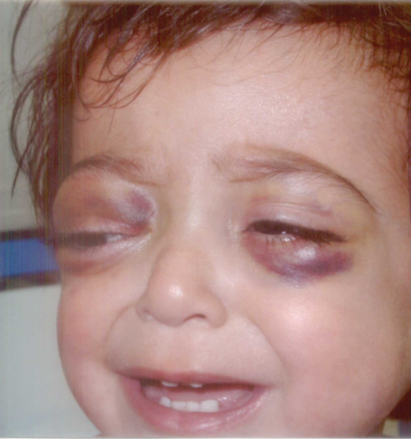
Raccoon Eyes


A I5-month-old girl was admitted to hospital with the complaints of bruising around the eyes for 10 days. She had one month history of abdominal pain and accompanied by fever and vomiting for one week. Physical examination demonstrated pallor, periorbital ecchymoses (raccoon eyes) and bilaterally mild proptosis (Fig. 1). She also had a left sided abdominal mass, which was 5 × 8 cm in diameter. The laboratory investigation revealed hemoglobin level 6g/dL, white cell count 10,000/mm3 and platelet count 34,000/mm3. Urinary vanillymandelic acid level was high. Bilateral bone marrow aspiration revealed infiltration with neuroblasts. Abdominal CT showed a left supra-renal mass (5 × 7 cm in diameter), which was diagnosed as a neuroblastoma on histopathologic examination.

Fig. 1. Bilateral periorbital ecchymosis (raccoon eyes) and mild proptosis.

The metastatic involvement of the periorbital tissues. has been described and the resultant proptosis and orbital ecchymosis has been given the tag of ‘raccoon eyes’. Orbital metastases can be found in up to 20% of children with stage IV neuroblastoma. The raccoon eyes appearance associated with neuroblastoma is probably related to obstruction of the palpebral vessels by tumor tissue in and around the orbis. There are a multitude of differential diagnoses for the presentation of periorbital edema and ecchymosis, e.g., child abuse or trauma, infection of the soft tissues associated with a spreading dental infection and an allergic reaction. Other systemic causes to be considered include myxoedema, other neoplasias such as lymphoma or haematological coagulopathies such as haemophilia.

Ali Bay,
A. Faik Oner,

Yuzuncu Yil University,
Faculty of Medicine,
Department of Pediatrics,
Van/Turkey.


Home

Past Issue

About IP

About IAP

Feedback

Links

 Author Info.

  Subscription