|
|
Images in Clinical Practice Indian Pediatrics 2003; 40:904-905 |
||
|
Addison’s Disease |
||
|
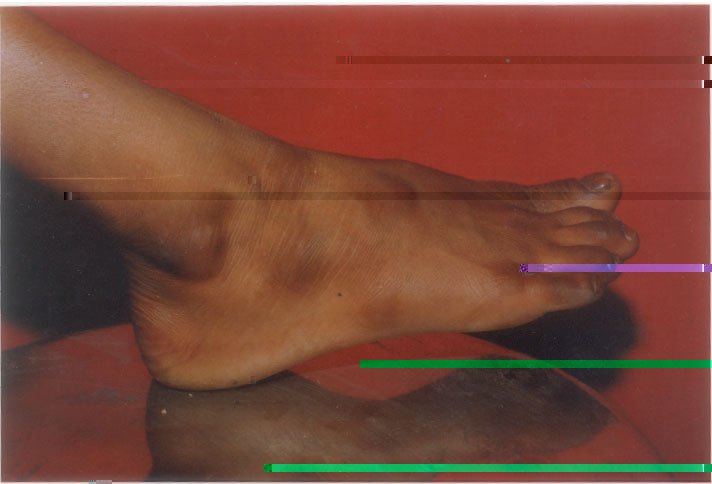
A 8-year-old boy presented episodes of early morning vomiting for 3 months. These episodes were relieved only by parenteral fluid therapy and antiemetics. On examination he was lethargic with a weight of 16 kg and a height of 125 cm. There were areas of increased pigmentation at pressure points on both his legs (Fig. 1). Blood pressure and systemic examination were normal. These clinical features suggested a diagnosis of Addison’s disease. The diagnosis was confirmed by serum cortisol (4.74 µg/dL, normal range 5 to 25 µg/dL) and serum ACTH (7934 pg/ml, normal range 0 to 37 pg/ml estimations. The child treated with was intravenous hydrocortisone initially for 3 days in a dose of 10 mg every 8 hours and later put on alternate day prednisolone in a morning dose of 10 mg/day.
Addison’s disease, a rare pediatric illness, with a prevalence of 1:100,000 can affect any age group and the presentation is with muscular weakness, fatiguability, weight loss, low blood pressure and darkening of skin specially in exposed parts of the body. Hyperpigmentation of skin is typically seen on mucous membranes scars, skin folds, and pressure points over knees and ankles. Similar areas of hyperpigmentation may also be seen in freckles; lentigines, and cafe au lait spots. Localised hyperpigmentation over lips and mucous membranes may be seen in Peutz Jeghers syndrome. N.P. Chhangani, |
![]()