|
|
|
Indian Pediatr 2017;54: 882 -884 |
 |
Hepatic Visceral Larva
Migrans Causing Hepatic Venous Thrombosis and Prolonged Fever
|
|
Jaswinder Kaur, Anand Gupta and Nishant Wadhwa
From Division of Pediatric Gastroenterology, Hepatology and
Nutrition, Sir Ganga Ram Hospital, New Delhi, India.
Correspondence to: Dr Nishant Wadhwa, Senior Consultant and Chief,
Division of Pediatric Gastroenterology, Hepatology and Nutrition, Sir
Ganga Ram Hospital, New Delhi, India.
Email:
[email protected]
Received: November 12, 2016;
Initial Review: April 07, 2017;
Accepted: July 27, 2017.
|
Background: Visceral larva migrans may present
with systemic symptoms such as fever, hepatomegaly, pneumonitis or
ocular symptoms. Case characteristics: A 7-year-old girl with
fever, pain abdomen and persistent eosinophilia. Imaging and
histopathology were suggestive of visceral larva migrans. Message:
The diagnosis of visceral larva migrans is often delayed since similar
symptoms of fever, hepatomegaly and peripheral eosinophilia occur in
more common and identifiable tropical parasitic and non-parasitic
diseases.
Keywords: Budd chiari syndrome, Fever of unknown origin, Liver
abscess, Toxocara canis.
|
|
V
isceral Larva Migrans (VLM) is a systemic
zoonotic parasitic disease due to migration of second stage larva of
Toxocara canis or Toxocara catis through viscera of
human beings. Poor hygiene, contact with dogs and geophagia increases
the risk of toxocariasis. Young adults and children who are in close
contact with animals are at a higher risk [1]. In VLM, the migrating
larva incites inflammation of the organs. The majority of patients are
asymptomatic and the infection resolves spontaneously. However in some,
the inflammation is severe resulting in morbidity if not treated
promptly. The clinical manifestations depend on the location of the
larvae, intensity of infection, duration of disease and host immune
response. VLM is under-diagnosed since there are no specific
symptomatology, ova is not identified in the feces and the findings on
imaging are very subtle. We present a 7-year-old girl initially treated
as pyogenic liver abscess and later diagnosed as hepatic VLM and
effectively managed with albendazole.
Case Report
A 7-year-old girl, resident of Jhansi, Uttar Pradesh,
presented with to us history of persistent high grade fever with chills
and intermittent pain abdomen for past six months. The pain was
localized to right hypochondrium with no aggravating or relieving
factors. There was no history of jaundice, black stools, bleeding per
rectum, passing worms in stool, loss of weight or pica. There were no
pets at home but there were many stray dogs in the neighbourhood.
Ultrasonography (USG) and Magnetic resonance imaging (MRI) done at
referring hospitals were suggestive of liver abscess, and she had
received multiple courses of antibiotics and metronidazole. At admission
in our health care facility, child was sick looking, febrile with mild
pallor. Examination of the abdomen revealed mild distension with
non-tender and firm hepatomegaly. There was no free fluid. Growth,
development, and rest of the systemic examination were normal. Based on
clinical features and available investigations, we considered
possibilities of pyogenic/amebic liver abscess, sepsis or enteric fever
and treated her with broad spectrum antibiotics. Investigations revealed
anemia (Hb 9 g/dL), leucocytosis (TLC: 17.9 × 10 9/L),
eosinophilia (41%), raised absolute eosinophil count (7.4 × 10⁹/L)
and normal liver function tests. Blood culture and Widal test were
negative. USG showed an irregular non-liquified necrotic area (103 cc)
with echogenic wall and multiple other echogenic lesions in right lobe
of liver. Middle hepatic vein was thrombosed with a tubular worm like
structure seen within it suggestive of VLM with secondary Budd Chiari
syndrome. In addition, a small echogenic partial thrombus was seen in
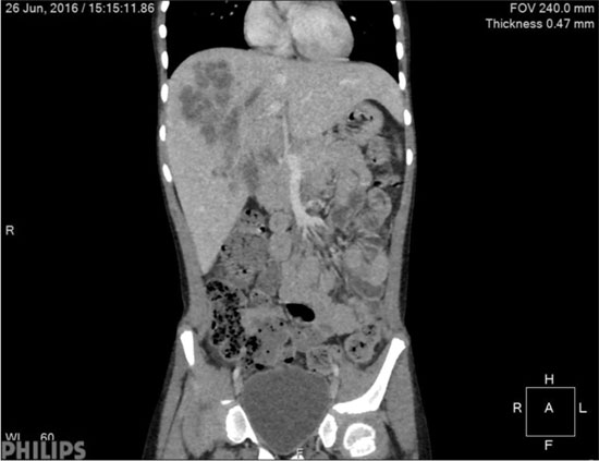
the left portal vein. Contrast enhanced computed tomography (CECT) of
abdomen showed large well-defined hypodense lesion with multiple
internal septations in liver, showing enhancement in the periphery and
septa on the portal phase with evidence of middle hepatic vein
thrombosis (Fig. 1). A liver biopsy was done which showed
presence of microabscesses composed of eosinophils, which were
also found in the sinusoidal spaces (Web Fig. 1).
No parasites were identified. The biopsy findings corroborated the
radiological findings and a provisional diagnosis of VLM was made.
Toxocara serology was not available at our centre and could not be sent
to referral centers due to financial constraints. Child was treated with
albendazole 400 mg twice daily along with prednisolone 2 mg/kg/day. She
improved dramatically and became afebrile within 48 hours of therapy.
Prednisolone was stopped and she was discharged after 10 days.
Albendazole was continued for 6 weeks as there was recurrence of fever
in spite of considerable resolution of hepatic lesions at the 3rd week.
After 6 weeks of therapy, USG showed minimal hepatic lesions, and
eosinophil count had reduced (AEC 1 × 10⁹/L).
 |
|
Fig. 1 CECT abdomen showing large
well defined clustered hypo-dense lesion with multiple internal
septations in liver with enhancement in the periphery and the
septa on the portal phase.
|
Discussion
In the present case, there were no pets at home but
there were plenty of stray dogs in the neighbourhood. The prevalence
rates of Toxocara eggs in soil samples in India is reported as 12 % [2].
The larva hatch, penetrate the intestinal wall and travel via the portal
vein to reach various organs. Liver is the most common organ to be
involved due to portal venous drainage and other involved sites are
lungs, heart, eyes and brain. During migration, larva excrete large
amount of glycosylated proteins which induce strong immune response
leading to eosinophilia and granulomatous inflammation [3]. Most cases
are asymptomatic and clinical manifestations depend upon the
localization, intensity and chronicity of the infection. As in the
present case, the child had been symptomatic for 6 months and there was
extensive liver involvement with venous thrombosis. There were no
clinical features of Budd Chiari syndrome as only single hepatic vein
and a branch of portal vein were blocked. The classic presentation of
VLM includes fever, hepatomegaly and eosinophilia as was seen in the
present case. Pulmonary involvement may lead to cough, wheeze or
pneumonia and neurological involvement may present with headache,
seizures or loss of consciousness [4]. The usual laboratory findings
include leucocytosis, marked eosinophilia (20% to 70%), raised absolute
eosinophil count raised IgE and hypergammaglobulinemia [5]. Imaging
forms an important role in diagnosis. As in our patient, eosinophilia
pointed towards parasitic infestation but VLM was thought of due to
suggestive USG and CT findings. CECT in hepatic toxocariasis usually
shows multiple, small, ill-defined, coalescing, low-attenuation nodules,
which are best appreciated on the portal venous phase. MRI findings
include hypointense lesions on T1 weighted sequence and hyperintense on
T2W with peripheral wall enhancement on contrast enhanced T1W images
associated with restriction of these lesions on Apparent Diffusion
Coefficient (ADC) maps corresponding to diffusion images [6]. The
resultant lesion on pathology is marked eosinophilic infilterates also
called eosinophilic abscess or granuloma as was found in our patient.
Toxocara larvae are rarely found on biopsy [7]. ELISA is the standard
serologic test to diagnose toxocariasis [5]. Though these tests are
available in India, the cost was a limiting factor in this child.
Anti-helminthic drugs form the mainstay of treatment
of VLM. Albendazole is given in a dose of 400 mg twice daily and
duration of the treatment depends upon the intensity of the infection
[8]. In our patient as there was extensive involvement, she received
albendazole for a total of six weeks. Corticosteroids and antihistamines
are often used to reduce the inflammation and prevent hypersensitivity.
In some cases, there may be no response to the treatment and surgical
excision may be needed [9]. VLM is often underdiagnosed due to low index
of suspicion and non-availability of the diagnostic methods. VLM may be
a cause of prolonged febrile illness and should be suspected in every
febrile patient with hepatic involvement and persistent eosinophilia.
Contributors: JK: collected the clinical details
and reviewed the literature; AG: managed the patient; NW: supervised the
management. All authors were involved in drafting the manuscript.
Funding: None; Competing interest: None
stated.
References
1. Hossack J, Ricketts P, Te HS, Hart J. A case of
adult hepatic toxocariasis. Nat Clin Pract Gastroenterol Hepatol.
2008;5:344-8.
2. Sudhakar NR, Samanta S, Sahu S, Raina OK, Gupta
SC, Madhu DN, et al. Prevalence of Toxocara species eggs in soil
samples of public health importance in and around Bareilly, Uttar
Pradesh, India. Vet World. 2013;6:87-90
3. Gutierrez Y. Diagnostic Pathology of Parasitic
Infections with Clinical Correlations. New York: Oxford University
Press; 2000.
4. Altcheh J, Nallar M, Conca M, Biancardi M, Freilij
H. Toxocariasis: clinical and laboratory features in 54 patients. Ann
Pediatr (Barc). 2003;58:425-31.
5. Luzna-Lyskov A, Andrzejewska I, Lesicka U,
Szewczyk-Kramska B, Luty T, Pawlowski ZS. Clinical interpretation of
eosinophilia and ELISA values (OD) in toxocarosis. Acta Parasitologica.
2000;45:35-9.
6. Laroia ST, Rastogi A, Sarin S. Case series of
visceral larva migrans in the liver: CT and MRI findings. Int J Case Rep
Images. 2012;3:7-12.
7. Kayes SG. Human toxocariasis and the visceral
larva migrans syndrome: correlative immunopathology. Chem Immunol.
1997;66:99-124.
8. Bhatia V, Sarin SK. Hepatic visceral larva migrans:
evolution of the lesion, diagnosis, and role of high-dose albendazole
therapy. Am J Gastroenterol. 1994;89:624-7.
9. Caumes E. Treatment of cutaneous larva migrans and Toxocara
infection. Fundam Clin Pharmacol. 2003;17:213-6.
|
|
|
 |
|
