|
|
|
Indian Pediatr 2016;53: 931-932 |
 |
Primary Epidural and Paraspinal
Rhabdomyosarcoma in a Child
|
|
*Sushil Kumar and
#Amit Garg
Departments of *Neurosurgery and #Radiology,
St. Stephens Hospital, New Delhi, India.
Email:
[email protected]
|
|
Rhabdomyosarcoma is the most common mesenchymal malignant tumor in
children but primary paravertebral location with spinal cord compression
is rare. A 2-year-old girl presented with swelling in upper back of two
month duration and progressive weakness of both lower limbs of 1½-month
duration. On examination, she had spastic paraparesis and a diffuse
swelling at the level of D
4-5,
firm to soft in consistency with ill-defined margins and mild
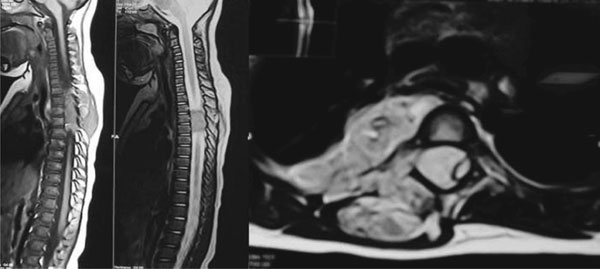
tenderness. Magnetic resonance imaging revealed a dumbbell shaped mass
at D3-5 level with extension
into chest wall and paraspinal area. The mass was isointense to spinal
cord on T1WI and hyperintense on T2WI (Fig. 1).
 |
|
(a)
(b)
(C)
Fig. 1 (a) Sagittal T1WI MRI showing
isointense epidural and paraspinal mass at D3-D5 level; (b)
Sagittal T2WI MRI showing hyperintense mass; (c) Axial T2WI
showed T2 hyperintense dumbell-shaped epidural mass involving
adjoining chest wall with severely compressed cord displaced to
the left.
|
D3-5
laminectomy revealed a fleshy, vascular mass with areas of hemorrhage.
Tumor from epidural and paraspinal area was removed completely.
Histopathology and immunohistochemistry confirmed the diagnosis of
alveolar rhabdomyosarcoma. Tumor cells expressed vimentin and desmin.
Patient showed rapid improvement in motor power in immediate
post-operative period and was given 12 cycles of chemotherapy (Vincristine,
Adriamycin , Cyclophosphamide, Mesna) and 41 Gy of radiotherapy.
Positron Emission Tomography scan in the follow up period revealed no
recurrence of the tumor and patient is symptom-free for the last three
years.
Primary spinal epidural rhabdomyosarcoma is an
extremely rare tumor and only few cases have been reported [1-4].
Treatment includes combination of surgery,
chemotherapy and radiotherapy. Prognosis depends upon the age of
patient, extent of the tumor, tumor histology, and presence of
metastasis. When an epidural spinal mass with nonspecific imaging
findings is found, rhabdomyo-sarcoma should be included in the
differential diagnosis. Follow-up imaging is important to monitor tumor
regression during or after completion of chemotherapy and radiotherapy,
and to detect tumor recurrence or metastasis.
References
1. Khalatbari MR, Jalaeikhoo H, Hamidi M, Moharamzad
Y. Primary spinal epidural rhabdomyosarcoma: A case report and review of
the literature. Childs Nerv Syst. 2012; 28:1977-80.
2. Rumboldt Z, Jednacak H, Talan-Hraniloviæ J, Kalousek
V. Spinal epidural rhabdomyosarcoma. Acta Neurochir
(Wien). 2004;146:195-7.
3. Salam S, Ganiou K, Idrissi A, Karkouri M, Aksim M,
Ouzidane L. Paravertebral rhabdomyosarcoma: Rare etiology of spinal cord
compression. African J Neurol Sci. 2010;29:77-82.
4. Yadav P, Gujrati. A, Buch A. Paravertebral and
epidural sarcoma with spinal cord compression in a child: Case report
and review of the literature. Medical Journal of DY Patil University.
2015;8:520-4.
|
|
|
 |
|
