|
|
|
Indian Pediatr 2015;52:
889-890 |
 |
Adult Form of Scimitar Syndrome Presenting as
Severe Pulmonary Hypertension in a Child
|
|
Sivasambo Kalpana, Sarath Balaji B, Selladurai
Elilarasi and *Neville AG
Solomon
From Department of Pulmonology, Institute of Child
Health and Hospital for Children; and *Department of Pediatric Cardiac
Surgery, Apollo Children’s Hospital; Chennai, India.
Correspondence to: Dr Sivasambo Kalpana, Department
of Pulmonology, Institute of Child Health and Hospital for Children,
Halls road, Egmore, Chennai.
Email: [email protected]
Received: February 21, 2015;
Initial review: April 14, 2015;
Accepted: May 29, 2015.
|
|
Background: Scimitar syndrome is a rare association of congenital
cardiopulmonary anomalies; the adult form is not usually is associated
with pulmonary hypertension. Case characteristics: 6-year-old
girl with recurrent episodes of cough and breathlessness, along with
features of right heart enlargement. Computed tomography angiogram
revealed right pulmonary veins draining into inferior vena cava with
dextroposition of heart. Outcome: Successfully managed with
surgical correction. Message: Scimitar syndrome should be
considered in any child with unexplained pulmonary hypertension and
dextroposed heart.
Keywords: Congestive cardiac failure,
Dextroposition, Pulmonary vein anomalies.
|
|
Scimitar syndrome has a varied presentation from
an asymptomatic state to severe pulmonary hypertension and/or heart
failure. Pulmonary arterial hypertension (PAH) is an uncommon
presentation of this syndrome beyond infancy.
Case Report
A 6-year-old developmentally normal girl was referred
to our department for evaluation of uncontrolled wheeze. There was
history of recurrent episodes of breathlessness and cough since 2 years
of age and exercise intolerance since 4 years of age. She had been
treated with intermittent salbutamol nebulizations and oral antibiotics,
with partial response. On admission, the child had no pallor, cyanosis
or clubbing. Significant respiratory distress was present with bilateral
severe wheeze; oxygen saturation in room air was 92 % that improved to
94% with 100% oxygen. There was a bulge on right side of chest wall with
heart sounds heard well on the right; P2 was loud and heart rate was
110/min. There was no murmur. With the above clinical picture, a
provisional diagnosis of Idiopathic pulmonary arterial hypertension with
congestive cardiac failure was made.
Chest X-ray showed right sided cardiomegaly
with pulmonary congestion. Electrocardiogram (ECG) demonstrated right
axis deviation with right atrial enlargement. Echocardiogram revealed
severe pulmonary hypertension (mean pulmonary artery systolic pressure
58 mmHg), and dilated right atrium and ventricle with a stretched patent
foramen ovale. Serum N-terminal prohormone of brain natriuretic peptide
was slightly raised to 144 pg/mL (normal 125 pg/mL). Computed tomography
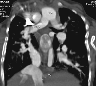
(CT) angiogram demonstrated that right pulmonary veins joined to form a
single vein which was draining into the inferior vena cava (IVC) with
dextroposition of heart (Fig. 1). The drainage of the left
pulmonary veins was normal. A persistent left superior vena cava (SVC)
and atrial septal defect (ASD) were also demonstrated. The diagnosis of
adult form of Scimitar syndrome presenting with severe PAH was made,
based on the clinical presentation and CT angiogram findings. The child
underwent intracardiac repair in view of the severe symptoms and Pah,
using right posterolateral thoracotomy. The right pulmonary vein was
dissected and reimplanted onto the left atrium (LA) using a autologous
pericardial patch, and closure of ASD was done. Intra-and post-
operative course of the child was uneventful.
 |
|
Fig. 1 CT angiogram showing right
pulmonary veins joining to form a single vein which is draining
into the inferioer vna cava.
|
Discussion
The hallmark of scimitar syndrome is an anomalous
right pulmonary vein that drains part or the entire right lung into the
IVC. Associated anomalies include hypoplasia of the right lung,
dextroposition of the heart, hypoplasia of the right pulmonary artery
and anomalous systemic arterial supply from the aorta to the right lung.
It has three main forms: an infantile form with severe symptoms and
pulmonary hypertension, an adult form distinguished by being
asymptomatic in infancy, and a third form with associated congenital
cardiac anomalies [1]. The diagnosis may be difficult, especially in
children and young adults with concomitant congenital heart lesions. CT
angiogram helped to clinch the diagnosis in this child and appears to be
an essential investigation in all children with unexplained pulmonary
hypertension, as echo-cardiography may miss the diagnosis (as in this
case) [2].
Pulmonary artery hypertension is seen mostly in
infants with associated congenital heart malformations or with an
anomalous large systemic arterial supply to the right lung [3]. A less
common cause of pulmonary artery hypertension is the presence of
Scimitar vein stenosis. Although our case did not have significant
hypoplasia or systemic arterial supply to the right lung, intraoperative
findings demonstrated significant stenosis at entry of the scimitar vein
into the IVC. This finding may explain the early presentation of this
child with severe pulmonary hypertension [4]. Unrelenting pulmonary
hypertension can cause irreversible damage to the pulmonary vascular bed
and lead to eventual right heart failure as in this child. Associated
cardiac anomalies frequently determine the clinical course of these
patients. About 70% of patients with scimitar syndrome have an
associated ASD. The presence of persistent left SVC, as in our patient,
has been described as a rare association [5].
Indication for surgical correction is based on the
severity of symptoms, pulmonary over-circulation, right ventricular
dilatation and concomitant cardiac lesions [6]. The surgical repair of
Scimitar syndrome consists of redirecting the pulmonary venous drainage
into the LA, either baffling the anomalous drainage into the LA via a
tunnel or transecting the "scimitar drainage" near its entrance into the
IVC and then reimplanting it directly into the LA. The majority of
patients show good clinical outcome on follow-up. Scimitar vein stenosis
may occur in about 15.5% at 10 years follow- up, irrespective of
surgical approach [7].
Contributors: KS: case management and writing the
manuscript; SBB and SE : case management and critical appraisal of
manuscript; NAGS: in case management. All authors approved the final
manuscript.
Funding: None; Competing interests:
None stated.
References
1. Dupuis C, Charaf LA, Brevie‘re GM, Abou P,
Re´my-Jardin M, Helmius G. The "adult" form of the scimitar syndrome. Am
J Cardiol. 1992;70:502-7.
2. El-Medany S, El-Noueam K, Sakr A. Scimitar
syndrome: MDCT imaging revisited. Egypt J Radiol Nucl Med.
2011;42:381-7.
3. Huddleston CB, Exil V, Canter CE, Mendeloff EN.
Scimitar syndrome presenting in infancy. Ann Thorac Surg. 1999;67:154-9.
4. Dupuis C, Rey C, Godart F, Vliers A, Gronnier P.
Scimitar syndrome complicated by stenosis of the right pulmonary vein:
apropos of 4 cases. Arch Mal Coeur Vaiss. 1994;87:607-13.
5. Sun J, Zhang S, Jiang D, Yang G. Scimitar syndrome
with the left persistent superior vena cava. Surg Radiol Anat.
2009;31:307-9.
6. Brink J, Yong MS, d’Udekem Y, Weintraub RG,
Brizard CP, Konstantinov IE. Surgery for scimitar syndrome: the
Melbourne experience.Interact CardioVasc Thorac Sur. 2015;20:31-4.
7. Vida VL, Padalino MA, Boccuzzo G, Tarja E,
Berggren H, Carrel T, et al. Scimitar syndrome: a European
Congenital Heart Surgeons Association multicentric study. Circulation.
2010;122:1159-66.
|
|
|
 |
|
