|
|
|
Indian Pediatr 2013;50: 982 |
 |
Black Heel (Talon Noir) Associated with a
Viral Exanthem
|
|
Kabir Sardana and *Vivek Sagar
Department of Dermatology, Maulana Azad Medical
College and Lok Nayak Hospital, New Delhi, India
*Department of Dermatology, ESI Model hospital, Sector
-9A.Gurgaon, Haryana.
Email: [email protected]
|
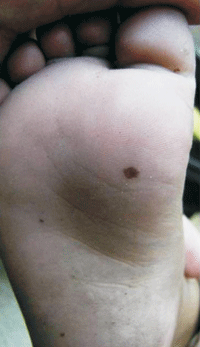
A 6-year-old child was referred for a suspected viral
exanthem associated with URTI. On examination the trunk
showed a maculopapular rash while the sole showed multiple
black "spots" which were present for 7 days. A diagnosis of
Talon noir was confirmed by paring the lesion with a No 15
blade which removed the superficial layer of the stratum
corneum, and revealed puncta of black pigment of
extravasated red cells.
 |
|
Fig. 1 Black macules with
irregular margins on the sole
|
This condition is normally seen as a
post-traumatic intraepidermal hemorrhage seen most often in
basketball players and is also known as "calcaneal petechiae".
The shearing forces rupture papillary dermal blood vessels,
with subsequent leakage of blood into the epidermis. The
major differentials include cutaneous melanoma, lentigines,
traumatic tattoo, verruca vulgaris and corn. Melanoma,
lentigines and traumatic tattoo cannot be removed by paring.
Dermoscopy, if available can distinguish Talon noir from
melanoma. Plantar warts are painful and bleed on paring
while corn do not bleed. In our patient probably the viral
infection predisposed to capillary fragility which in
association with the normal shearing force on the sole led
to the disorder. The child used to walk barefoot even before
the viral exanthema without any history of a similar lesion
in the past. Vitamin C 500 mg once a day lead to resolution
within 7 days. The disorder can also resolve spontaneously.
|
|
|
 |
|

