A 12-year-old girl presented with multiple, asymptomatic,
annular lesions over right side of upper chest and right
forearm for last 5 years. Initially, few small brown
coloured papules appeared over the chest which gradually
increased in number and size to attain the present status.
The lesions were asymptomatic from the beginning. There was
no history of similar episodes in the past or in the family,
and no history of any skin lesions at birth. On examination,
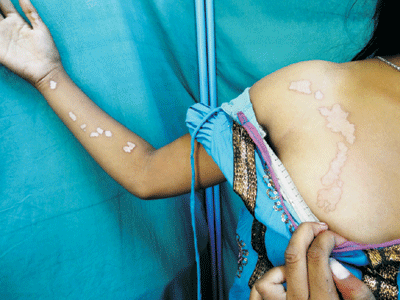
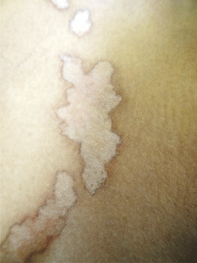
multiple skin coloured annular plaques (1.5-2 cms × 3-7 cms
in size) with a well-demarcated raised, thready margin with
central hypopigmentation and atrophy were found. They were
arranged in a linear configuration and unilaterally over
right side of upper chest and right forearm (Fig.1
and 2). The lesions had a tendency of peripheral
extension and central clearing. Mucosa, scalp and nails were
spared. Biopsy was done from one of the lesions and on
histopathological examination, hyperkeratosis, cornoid
lamella with perivascular dermal infiltrates were seen and
central part of the lesion showed atrophy. Biopsy findings
confirmed it to be a case of "unilateral linear
porokeratosis". She has been prescribed topical retinoids
and she is under regular follow-up because this variant of
porokeratosis is highly prone to develop malignancy.
 |
|
Fig. 1 Linear arrangement
of multiple annular plaques.
|
 |
|
Fig. 2 Skin lesions showing
demarcated raised irregular margins with central
hypopigmentation.
|
Porokeratosis is a disorder of
keratinisation, characterised by hyperkeratotic papules or
plaques surrounded by a thready elevated border that expands
centrifugally. Most cases are sporadic. Pathogenesis is
unclear. Seven varities are described. Linear type presents
in early childhood and has highest potential for developing
squamous cell carcinoma. Histopathological examination gives
the definitive diagnosis. The differential diagnoses to be
considered are inflammatory linear verrucous epidermal nevus
(lesions since birth, erythema, scaling, itching present),
stage IV of incontinentia pigmenti (earlier age of onset,
preceded by vesicular, verrucous, hyperpigmented stage,
associated CNS, dental and ocular defect), linear lichen
planus (hyperkeratotic, violaceous, pruritic papule and
plaque). On histopathology none of them shows cornoid
lamella. Topical 5 fluorouracil, topical calcipotriol,
topical retinoid, cryotherapy and surgical excision have
been tried with various degree of success.


