|
|
Review Article Indian Pediatrics 2008; 45:829-837 |
|||||||||||||||||||||||||||||||||||||||||||||||||||||||||||||||||||||||||||||||||||||||||||||||||||||||||||||||||||||||||||||||||||||||||||||||||||||||||||||||||||||||||||||||||||||||||||||||||||||||||||||||
|
Evaluation and Management of Photosensitivity in Children |
|||||||||||||||||||||||||||||||||||||||||||||||||||||||||||||||||||||||||||||||||||||||||||||||||||||||||||||||||||||||||||||||||||||||||||||||||||||||||||||||||||||||||||||||||||||||||||||||||||||||||||||||
|
From the Department of Dermatology, Bombay Hospital and Medical Research Center, Mumbai; and *Department of Pediatrics, Jagjivan Ram Hospital, Mumbai Central, Mumbai 400 008, India. Correspondence to: Dr. Rajesh Kumar, 208, 2 nd Floor, New Wing, Bombay Hospital and Medical Research Center, 12, New Marine lines, Mumbai 400020, India E mail: [email protected]
Photosensitive disorders in children are not uncommon in our country. It is often difficult to establish the diagnosis, either due to confusing presentation or ignorance by the parents. We review the presentation and management of photosensitive disorders in children. Detailed history and thorough clinical examination is essential in arriving at a particular diagnosis. Photosensitive disorders in children are idiopathic, or related to nutritional, genetic, metabolic or connective tissue diseases. Most photosensitive disorders begin in infancy or childhood and persist for invariable period during adulthood. However, their severity decreases with age. As there is no definitive treatment in most of the cases, the only treatment is prevention from further exposure to sunlight. Our aim should be to minimize the photo damage which can be achieved by adopting different methods of photoprotection. Photoprotection can be achieved by using adequate sunscreens, proper clothing and avoidance of sun exposure. It is recommended that these children and their parents should be adequately counseled about the disease and different methods of photoprotection. This article describes how to diagnose photosensitive disorders in children and its management. Key words: Children, Photosensitivity. Definition Photosensitivity is commonly defined as an abnormal reaction of the skin to sunlight and other sources of UV light(1). A child is said to be photosensitive, if he or she develops reddening and blistering of the skin after exposure to sunlight or ultraviolet light(1-4). In other words, a photosensitive child may develop symptoms even after brief exposure to sun. Phototoxicity vs. Photoallergy Photosensitive disorders are broadly classified as either phototoxic or photoallergic(5-6). It is often difficult to differentiate between the two, however they can be differentiated by certain signs and symptoms. For phototoxic reaction to occur, prior sensitization is not required(6). The onset is acute and can lead to blister formation. The lesions are angry looking and subside if there is no further exposure. Phototoxic reactions are usually caused by drugs. The list includes several common antibiotics-quinolones, sulfonamides, and tetracyclines; diuretics; major tranquilizers; oral antidiabetic medication; and cancer medicines(6). In contrast, photoallergic reactions are a delayed hyper-sensitivity reaction characterized by eczematous skin lesions. These lesions appear gradually and persists for variable period of time. They are commonly due to sunscreens (most common) and fragrances used in different household products like perfumes and cosmetics(5,6). These reactions (phototoxic and photoallergic) get aggravated if the child has underlying photosensitive disorder(5). These disorders could be idiopathic, nuitritional, metabolic, genetic or due to systemic diseases like systemic lupus erythematosus (SLE)(2). Few conditions get aggravated after sun exosure like atopic dermatitis and pellagra. Clinical Features The majority of patients report a pruritic, burning or tingling sensation immediately after exposure to sun or UV light(1-7). The duration of exposure required to induce these symptoms depend upon the underlying disease and environmental conditions. Erythema (redness) and swelling on exposed part of the body (face, upper back, chest, extensor aspect of forearm) are common features in a child suffering from photosensitive disorder. In an acute episode apart from erythema (reddening), child develops blisters. With repeated exposure, child may develop butterfly erythema on face, persistent dermatitic plaques, telangiectasia and poikilodermatous changes (Table I). Specific clinical diagnosis in a particular case can be made by combination of signs and symptoms suggestive of a particular photosensitive disease. Photosensitive disorders in children are grouped into five categories namely idiopathic, nuitritional, metabolic, genetic and connective tissue diseases (Table II)(2). TABLE I Clinical Signs in a Photosensitive Child
* Triad of atrophy of skin, reticulate pigmentation, and telangiectasia. TABLE II List of Photosensitivity Disorders in Children
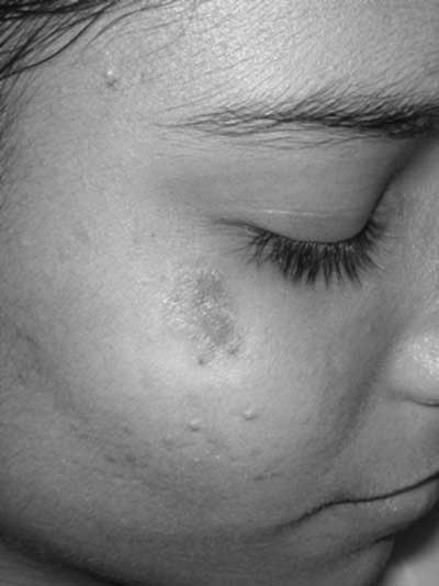
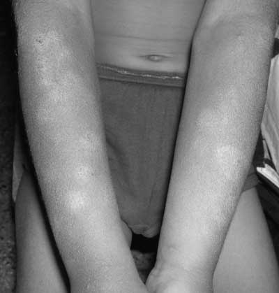
Idiopathic Photosensitive Disorders In this group of disorders, exact pathogenesis is not known. There is no systemic involvement as compared to metabolic and genetic photosensitive diseases. The diseases are polymorphous light eruption (PMLE), actinic prurigo and hydroa vacciniforme. Most common in our part is PMLE. Polymorphous light eruption (PMLE) is a common, idiopathic, inflammatory disorder affecting children of school going age(2,3). The affected child may have small itchy papules, papulovesicles, or large urticarial plaques occurring in areas of sun-exposure (Fig. 1 and 2)(8). These lesions are of different shapes and sizes (hence the name polymorphous). Typically, the lesions appear several hours to days after sun exposure and subside within 7-10 days, if there is no further exposure(8).
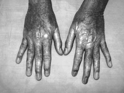
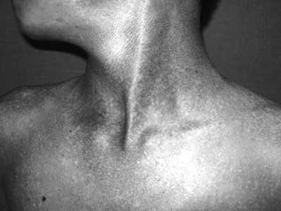
Actinic prurigo (AP) is a rare inflammatory photodermatosis which usually starts in childhood(2,9,10). It is more common in Caucasian population(9,10). Symptoms are similar to those of PMLE, but the lesions are concentrated on the face, especially around the lips (cheilitis)(9,10). Hydroa vacciniforme (HV) is a chronic photodermatosis that usually begins in childhood and resolves spontaneously by early adulthood(2,11). The lesions follow a distinct clinical pattern. After exposure to sunlight, child develops pruritus, erythema and swelling on exposed parts(11). Later they develop tender papules which become vesicular (hydroa) and tense(11). Gradually, it becomes umblicated and hemorrhagic and subsequently forms crust (seems to follow clinical course of chicken pox, hence it is named vacciniforme). The crust separates out in about a week leaving behind depressed chickenpox-like scars(11). Nutritional Deficiency Disorder Pellagra is due to inadequate dietary intake of niacin or its precursor (tryptophan)(12). It is usually seen in children, with malabsorption syndromes, corn as a staple diet and in adolescents with anorexia nervosa. Apart from systemic symptoms, child may develop dermatitic plaque confined to sun exposed sites- face, neck, dorsal surface of hands and feet. The dermatitic plaque is well defined, which subside with dusky brown-red discoloration. On the neck, the plaque appears as broad collarette of dermatitis known as Casal’s necklace. Measurement of the urinary excretion of N1- methylnicotinamide and/or pyridine is helpful in suspected cases(12). Treatment of pellagra consists of high-protein diet and nicotinic acid or nicotinamide. If good diet is maintained, complete recovery is the rule. Inborn Errors of Metabolism Congenital erythropoietic porphyria (CEP) or Günther’s disease manifests during infancy. It is a rare autosomal recessive disease characterized by cutaneous fragility, photosensitivity and having tendency to scarring and mutilation. Children develop photosensitivity (redness and burning) on exposed part of the body followed by blisters, which heal by scarring. Hypertrichosis, hemolytic anemia, progressive splenomegaly, photophobia, excessive tearing, keratoconjunctivitis, loss of eyelashes and ectropion may occur(13). The urine is dark and deciduous teeth are red in color (erthrodontia)(13). The disease is slowly progressive in nature and has mutilating tendency(2,13). Erythropoietic protoporphyria (EPP) is an autosomal dominant disorder characterized by photo-sensitivity, which is variable and unpredictable(2,14). Often the parents are unaware of their child’s photosensitivity. The age of onset is between 3-5 years. The child develops pain immediately after exposure to sunlight. The skin becomes yellowish, thickened and waxed over dorsa of hands and feet(14). Hartnup disease is a rare inborn error of metabolism with autosomal recessive inheritance characterized by pellagra like cutaneous eruptions, neurologic abnormalities and a specific amino-aciduria. Cerebellar ataxia is a prominent feature which manifests as unsteady, wide-based gait(15). Photosensitive Genetic Diseases These disorders are differentiated from each other on the basis of their clinical features and age of presentation (Table III). TABLE III Clinical Feature of Genetic Photosensitive Disorders
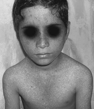
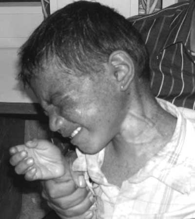
AR: autosomal recessive Xeroderma pigmentosum (XP) is characterized by photosensitivity, photo-damage, and cutaneous malignancies. The cells of patient with XP have defect in DNA repair(2,3,16). Generalized prog-ressive freckling and photophobia are common presenting feature (Figs. 3,4). Children with Cockayne’s syndrome look cachectic and dwarf(15,16). Weight is more affected than height, hence it is termed "cachectic dwarfism"(17,18). Freckling and cutaneous malignancies are not a feature of CS as compared to XP(17). Microcephaly and cachectic look give them "Mickey-mouse like" facies(3,17). Bloom syndrome (BS) is characterized by growth retardation, immunodeficiency, photo-sensitivity, telangiectasia and pigmentation (19,20). Kindler syndrome (KS) is characterized by acral blister formation in infancy and childhood, photosensitivity, generalized progressive poikilo-derma and cutaneous atrophy (Figs. 5,6)(21,22). Characteristically, the photosensitivity and blister formation improves with age. Children suffering from Rothmund-Thomson syndrome (RTS) may have transient photo-sensitivity, short stature, sparse hair, hypoplastic nails, cataract, hypogonadism and midface hypoplasia(23,24).
Connective Tissue Diseases The connective tissue diseases which manifests in childhood are lupus erythematosus (LE) and juvenile dermatomyositis (DM). Neonatal lupus erythe-matosus is a rare syndrome, comprising of transient skin lesions and / or congenital heart block(25,26). It is due to transplacental passage of maternal auto-antibodies to the ribonucleoproteins (RNPs), Ro-SSA, La-SSB, or U1RNP. The skin rash is either present at birth or evolves during first few months of life(25). The typical skin lesions are well-defined macular or slightly elevated annular (ring) plaques occurring predominantly on face, particularly the forehead, temples and upper cheeks(25). The rash is often misdiagnosed as tinea corporis or atopic dermatitis. The skin lesions in most cases disappear within first year with residual atrophy and telangiectasia. Childhood systemic lupus erythe-matosus presents with skin rash, arthritis and nonspecific constitutional symptoms. Affected child may develop discoid rash, malar rash and cutaneous ulceration. These lesions are very photo-sensitive(27,28). Juvenile dermatomyositis is a rare, chronic, multisystem inflammatory disorder, which manifests as skin rash on exposed parts and proximal muscle weakness. The other cutaneous manifestations are heliotrope rash, Gottron’s papules, and calcinosis cutis(29). Evaluation of a Child with Suspected Photosensitivity The first and foremost task is to establish a diagnosis that a given child is photosensitive. It is commonly seen in practice that parents seek consultation when there is no lesion. They may be advised to consult when new lesion appears. Photosensitive disorders must be differentiated from reactions due to excessive and prolonged exposure to sunlight, which is a normal response in any child. Photosensitivity should be suspected if a child develops erythema, swelling, intense pruritus after limited exposure to sunlight. Hence detailed history is very important while evaluating a child with suspected photosensitive disease(1,5,7). It helps in investigating an individual case. In history one should ask about the age of onset, exposure to potential photosensitizers, and seasonal variation(1). The child should be examined in a well lit room preferably in natural day light to look for extent of involvement. Examination should focus on the distribution of lesions, including area of sparing. In a photosensitive disorder, the upper eyelids, postauricular, area under the chin and lower lip, nasolabial folds, neck folds, volar aspect of wrist and the antecubital fossae tend to be spared. The morphology of the skin lesions is important in differentiating acute versus chronicity of photo-sensitive disorders. Urtricarial, papular, vesicular lesions indicate acute episode as compared to lichenified (thickened) lesions of chronicity. Those with childhood SLE and dermatomyositis require detailed clinical examination to look for any underlying systemic involvement i.e., hepatosplenomegaly, proximal muscle weakness and muscle tenderness. In a photosensitive disorder, diagnosis is usually made by history and clinical findings(1-5,30). Lesions can be reproduced by graded exposure to UV light as we do in phototesting or photo patch testing. However, it is difficult to perform phototesting and photopatch testing in children(3). These tests need motivation and cooperation which is not possible, especially in small children. When the suspected diagnosis is pellagra, measurement of the urinary excretion of N1- methylnicotinamide and/or pyridine is helpful(12). Biochemical investigations and Wood’s lamp examination are required in case of porphyrias. The urine of patients with porphyrias shows reddish pink fluorescence under Wood’s lamp. The diagnosis of Hartnup disease is based on the clinical picture and demonstration of specific amino acid (neutral amino acids like alanine, serine, threonine, valine etc.) excretion pattern. All patients with photosensitivity without definitive clinical diagnosis should be evaluated for antinuclear (ANA), anti-Ro (SSA), anti-La (SSB) antibodies and porphyrin levels(31,32). Skin biopsy and direct immunofluorescence is helpful in cases of SLE where there are deposition of immunoglobulin and complement at dermoepidermal junction(28). Management Most of the photosensitive diseases in children are either genetic or metabolic. As there is no cure, or few conditions improve with age, these children and their parents need detail counseling. The problem should be discussed repeatedly. The emphasis should be on sun avoidance and protection(33). Sun-avoidance: During the mid-part of the day, the sun’s rays strike the earth more directly and therefore, more UV penetration. Children should minimize or avoid outdoor activities between the peak hours of 10:00 a.m. to 4:00 p.m. Reflective surfaces such as sand, snow and water surface increase exposure to UV light. Snow skiing or sitting on a sandy beach exposes one to a lot of reflected UV light. A child’s UV exposure is significant even if they are sitting under an umbrella at the beach. Cloudy sky does not reduce UV substantially and provides only minimal photoprotection from UV rays(34,35). Photoprotection: The simplest and effective way of photo-protection is clothing that can effectively block UV rays(34,35). The thickness of the clothing is more important than color. The tighter the weaving of the cloth, better the protection. Thin, loosely woven cloths should be avoided as it offers little or no protection(35). Long sleeve shirt and long pants preferred over half sleeve shirt and shorts. When sun exposure is unavoidable, facial exposure can be minimized by using wide brimmed hat, umbrella and sunscreen. Sunscreens: Sunscreens are chemicals which give protection by absorbing and scattering UV light(33-35). They are available as creams, lotions or gels. The chemical content determines whether the sunscreen absorbs UVB, UVA or both. Chemicals that primarily absorb UVB include PABA (Para-amino benzoic acid), and its esters, cinnamates and salicylates. Padimate-O is the most widely used UVB absorber. Benzophenones and anthranilates absorb both UVB and UVA. Avobenzone (Parsol 1789) is an excellent UVA absorber(35). UVB sunscreens are rated according to their sun protective factor (SPF)(35). An SPF of 15 protects the skin so it takes 15 times longer to produce erythema (redness) than it would without the sunscreen. SPF varies from 1-50. While purchasing a sunscreen consider the following: the SPF number, the type of UV absorbed and its ability to remain effective after application(35,36). For Indian skin, SPF 15 or more is sufficient. Exercise, sweating, and swimming decreases a sunscreen’s effectiveness; it is advisable to apply "water resistant" sunscreens before these activities(35,36). A sunscreen is considered water-resistant if the SPF level is even effective after 40 minutes of water immersion. Till date, there is no universally accepted parameter to judge the efficacy of sunscreen for UVA protection(33-36). Sunscreens may produce side effects (contact dermatitis). It is always advisable that before applying a new sunscreen, first test the product on a small area of skin preferably on back of child to rule out sensitivity or allergy. If the problem of allergy persists, a patch test with probable antigen can help in identifying specific allergens(35,36). Proper instructions about the sunscreen should be given to parents and child. Ideally, a broad-spectrum sunscreen with a Sun Protection Factor (SPF) 15 or higher should be applied, 15 to 30 minutes before going outdoors. If a child is going to stay outdoors for more than three hours then it should be reapplied. Currently available sunscreen is effective up to 3 hours(34). Moreover, it should be stressed that no sunscreen can effectively blocks all part of ultraviolet spectrum(34-36). Certain drugs known to be photosensitizers such as tretinoin (Retino-A), hydrochlorothiazide, chlorpromazine, tetracycline, sulfa drugs, chlorpro-pamide and non-steroidal naproxen should be avoided. If it is necessary then the offending drug should be replaced by chemically unrelated compound(6,35). To summarize, avoid unnecessary sun exposure during peak hour (between 10 am to 4 pm); if not possible, wear tightly woven full sleeve shirt and trousers, wide brimmed hat, umbrella and apply broad spectrum sunscreen on exposed part. Corticosteroids: Corticosteroids, both topical and systemic can be given in case of acute photosensitivity to alleviate the symptoms. Topical steroids with mild to moderate potency should be given for a brief period (5-7 days). Fluticasone and mometasone furoate are moderately potent steroids with good safety profile (less atrophogenic) in children, can be used. Systemic steroids can be given for a brief period for faster recovery. Phototherapy and Photochemotherapy: Paradoxi-cally, graded exposure to sunlight or artificial UV rays exposure increases tolerance. The mechanisms by which ultraviolet B (UVB) or PUVA (oral ingestion of psoralen followed by ultraviolet A exposure) induce such tolerance are not completely understood(36). Pigmentation and skin thickening caused by UVB and UVA may be important factors in the protective effect. The method most commonly used as phototherapy in case of children is narrow band UVB, as PUVA is contraindicated(35,36). This modality of treatment can be tried after appropriate counseling and after consultation with a dermatologist Acknowledgment We are thankful to Dr Vijay Zawar, for providing some of the clinical photographs of his patients. Contributors: RK: concept and design, acquisition
of data and analysis; SK: Drafting and critical revision of the
manuscript.
| |||||||||||||||||||||||||||||||||||||||||||||||||||||||||||||||||||||||||||||||||||||||||||||||||||||||||||||||||||||||||||||||||||||||||||||||||||||||||||||||||||||||||||||||||||||||||||||||||||||||||||||||
![]()