|
|
Letters to the Editor Indian Pediatrics 2005; 42:1060-1062 |
|||
|
Meconeum Hydrocele Presenting asa Labial Mass |
|||
|
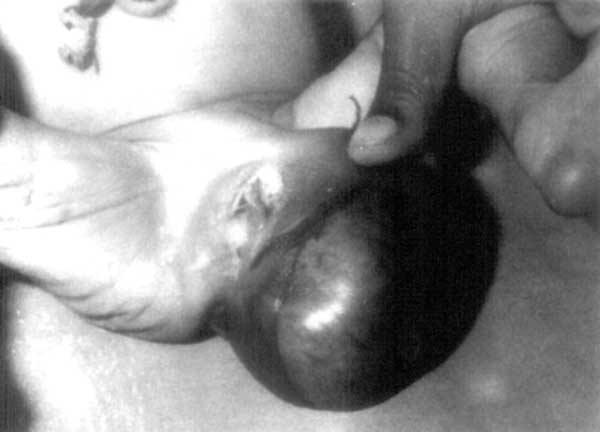
A one-day-old female baby presented to us at 7 hours after birth with a left labial mass of 7 × 4 × 4 cm, soft, cystic, normal colour of overlying stretched skin, no impulse on crying, noncompressible, positive trans-illumination in the upper 1/5th portion of the swelling and negative below that. The baby passed stool and urine normally on first day of life. On the second day of life, the swelling increased in size to 10 × 5 × 5 cm, turned yellowish in colour and there was impending perforation (Fig.1). The swelling ruptured spontaneously and the sloughed out skin was excised. The open cavity was dressed with povidone iodine and swelling never refilled. The baby passed stool normally. She was given intravenous antibiotics and discharged after 7 days. The wound healed up with granulation tissue. The baby is doing well at 3 months follow up.
Meconeum peritonitis results from extrusion of sterile meconeum into the peritoneal cavity after fetal perforation. The rich content of digestive enzymes of the meconeum leads to peritoneal irritation and inflammatory response characterized by foreign body granuloma formation and calcification. The majority of patients with meconeum peritonitis require surgical intervention but many have spontaneous resolution by sealing of perforation. The processus vaginalis forms by 6 months of gestation as an evagination of parietal peritoneum and descends ventral to the gubernaculum into scrotum or labium majus(2). In males, patency is maintained until birth or shortly thereafter with subsequent proximal obliteration leaving the residual tunica vaginalis. In females, the processus vaginalis is small and obliterated by 8 months of gestation. Autopsy studies have proved that the female gubernaculum or round ligament is different from the male in ending just outside the external ring(3). If patency persists, hydrocele of the canal of Nuck has been described and ultrasound has been suggested as the best mode of diagnosis(4). Delayed presentation may lead to formation of infection. Associated inguinal hernia has been reported in one-third of the cases(5). The differential diagnosis of meconeum hydrocele includes inguinal hernia, ectopic ovary, labial lipoma, fibroma, leiomyoma or hemangioma, round ligament sarcoma, inguinal lymphadenopathy and epidermal cyst(5). If there is a labioscrotal swelling, this remote possibility should always be kept in mind otherwise it may lead to severe inflammation and spontaneous rupture. Shilpa Sharma,
|
![]()