|
|
Letters to the Editor Indian Pediatrics 2005; 42:1056-1057 |
||
|
Idiopathic Pulmonary Hemosiderosis |
||
|
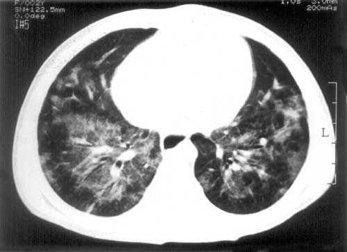
A 2-year-old female was admitted with apparently "non-resolving pneumonia" of one and a half months. A chest radiograph showed bilateral non-homogenous opacities, bronchoscopy did not yield a foreign body and bronchoalveolar lavage fluid showed no organism on Gram and Z-N stain. There was no past history of similar episodes, contact with tuberculosis, and family history of atopy. The child was immunized for age, weighed 10 kgs had fever, tachycardt.a, tachypnea and pallor, evidence of severe respiratory distress and congestive cardiac failure. The patient was thought to have acute onset pneumonia with an underlying lung pathology that needed further investigation and congestive cardiac failure, presumably due to nutritional anemia. Investigations revealed hemoglobin of 6.0 g/dL microcytosis and hypochromia, leucocytosis with predominant polymorphs. Arterial blood gas was suggestive of hyper-carbia and mild hypoxemia. Chest radiograph showed bilateral non-homogeneous opacities, with a small right sided pleural effusion, and no change in comparison to previous X-ray done 5 weeks earlier. She was treated with antibiotics and supportive measures including a packed cell transfusion and within twenty-four hours her condition had improved. Further investigations: absolute eosinophil count: 160/mm3, ELISA for anti-HIV antibody: negative, immunoglobulin profile: normal, anti-neutrophil cytoplasmic antibody (ANCA) negative, computerized tomography (CT) of chest: alveolar damage with hazy lung fields (Fig. 1). Thoracoscopic lung biopsy displayed multiple nodules of varying sizes on the lung surface, histopathologic examination exhibited alveolar spaces filled with macro-phages containing brown granular pigment, thickened alveolar septae with infiltration by mononuclear cells and partial atelectasis. Bronchoalveolar lavage fluid revealed abundant hemosiderin laden macrophages. This confirmed the diagnosis of pulmonary hemosiderosis and the patient was started on oral prednisolone. There was no evidence of myocardial involvement, valvular heart disease, pancreatic insufficiency, vasculitis or exposure to drugs. It is difficult to rule out cow’s milk allergy with certainty. On follow up two months later, the patient has had no recurrence of symptoms, her Hb is 12.0 g%, chest radiograph shows clearing of alveolar haziness though some reticular shadows remain.
The highly variable clinical presentation of PH often leads to a delay in its diagnosis. The classical triad of anemia, hemoptysis and pulmonary infiltrates is rare in children(4). Hemoptysis is unusual as children swallow blood stained sputum and alveolar bleeding does not readily gain access to central airways(1,3). Swallowed blood may lead to positive occult blood in stools, a misleading clue pointing to gastrointestinal blood loss as a cause for anemia. Our patient possibly had some pulmonary hemorrhage earlier, making her moderately symptomatic. Hemosiderin-laden macrophages in the lavage fluid in the initial bronchoscopy were not looked for. A recent episode of hemorrhage resulted in acute onset of severe anemia and congestive cardiac failure. It is difficult to diagnose IPH because nutritional iron deficiency anemia is common in our children, as is tuberculosis which may present with lung infiltrates not responding to routinely used antibiotics, pneumonia due to resistant organisms is on the rise, as is also HIV infection. A high index of suspicion for this disease in the relevant clinical setting will overcome the delay between the onset of illness and correct diagnosis. This may prevent antibiotic misuse and risk of death due to severe hemorrhage. Of note in context to IPH are recent reports of clustering of cases in infants in areas of United States. It has been recommended that recommendations from the working group for Investigation and surveillance of pulmonary hemosiderosis be followed for the diagnosis and investigation (5). Jyoti Sharma,
| ||
|
References | ||
|
|
![]()