1.gif (1892 bytes)

Letters to the Editor

Indian Pediatrics 2004; 41:1068-1069

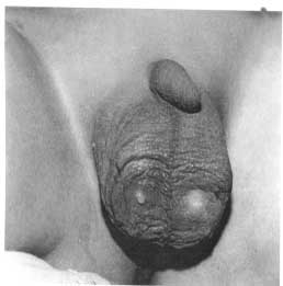
Posterior Urethral Valves with Down’s Syndrome presenting as Scrotal urinary Sinuses


A one and a half year old male was brought to us with bilateral anterior scrotal sinuses of one month duration (Fig. 1). The sinuses were draining clear urine. The child had earlier presented to another hospital with fever and bilateral inflamed scrotal swellings. This was thought to be a scrotal abscess and was drained. Initially it drained pus but subsequently scrotal sinuses were formed which drained clear urine intermittently. The child was dysmorphic and had features of Downs’ syndrome. He was well hydrated and the bladder was not palpable at the time of examination. The parents felt that the child had normal urinary stream. There was no history of antenatal oligohydramnios. Biochemical renal functions were normal. Ultrasound examination of the urinary tract did not reveal any abnormality. Micturating cystourethrogram showed a dilated posterior urethra suggestive of posterior urethral valves. Cystoscopy showed a hugely dilated posterior urethra. Large openings of the ejaculatory ducts were seen opening above the semilunar valves located below the Veru in the posterior urethra. Trans-urethral fulguration of valves was done at 5,7 and 12 O’clock position. The scrotal sinuses healed in a weeks’ time and the child is well and having good urinary stream after four years. His biochemical renal functions are normal. However there is mental retardation due to Down’s syndrome.

Fig. 1. Uninary leak from scrotal sinus.

Posterior urethral valves (PUV) present with varying severity and grades of obstruction to the urinary tract. This unusual presentation of PUV as non healing scrotal sinuses after epididymoorchitis has not been reported earlier. Association of PUV with Down’s syndrome is also rare though reported(1). Epididymoorchitis is caused by retrograde flow of infected urine through the ejaculatory ducts. The mechanism of retrograde flow of urinary stream into the testes may be explained as follows. The severe attenuation of prostatic tissue seen in congenital obstructions leads to loss of obliquity of the normal ejaculatory ducts as they enter the posterior urethra. Associated distal obstruction to the flow of urine may aid in the urethro-ejaculatory reflux of urine .If the urine is infected, this precipitates epididymoorchitis as it happened in the index case. However this reflux must be very unusual as epididymoorchitis is a rare association in PUV.

Epididymoorchitis is uncommon in children and is indicative of an underlying abnormality of the urinary tract, usually a pathological connection between the urinary system and the genital duct system or the bowel(2,3). Any pre-pubertal child with epididymitis merits a complete urological evaluation including urine culture, voiding cystourethrography and excretory urography. Surgically treatable conditions ectopic ureters opening into seminal ducts, ectopic vasal insertion into the bladder, and recto- urethral fistulas have been picked up on screening children of epididymoorchitis. Hence pediatricians encountering a child with epididymo-orchitis must exclude underlying surgical problems in the baby which are treatable(2,3).

Narasimhan KL,
Alka Gupta,

Department of Pediatric Surgery,
PGIMER, Chandigarh 160 012,
India.
E-mail: [email protected]

References

1. Kupfermann JC, Steward CL, Kaskel FJ, Fine RN. Posterior urethral valves in patient with Down syndrome. Pediatric Nephrol 1996; 10: 143-146.

2. Siegel A, Snyder H, Duckett JW: Epididymitis in infants and boys: underlying urogenital anomalies and efficacy of imaging modalities: J Urol 1987; 138: 1100-1103.

3. Umeyama T, Kawamura T, Hasegawa A, Ogawa O. Ectopic ureter presenting with epididymitis in childhood: A report of 5 cases. J Urol 1985; 134: 131-135.

Home

Past Issue

About IP

About IAP k

Feedback

Links

 Ahor Info.

  Subscription