|
|
Original Article Indian Pediatrics 2003; 40:951-957 |
||||||||||
| Vascular Rings: An important Cause of Persistent Respiratory Symptoms in Infants and Children | ||||||||||
|
Barium swallow, Double aortic arch,Vascular rings. Vascular rings are a group of congenital defects wherein the trachea and esophagus are completely encircled by vascular structures, which may or may not cause symptomatic compression. They form an important cause of unexplained respiratory symptoms in infants and young children(1). Early diagnosis and prompt surgical relief of airway obstruction can improve the patient’s symptoms dramatically. Although barium swallow studies are well known to identify the problem in a majority of patients, often this is not performed as part of initial workup, with consequent delay in diagnosis. This retrospective study of infants and children diagnosed to have symptomatic vascular rings was done to identify the types of anomalies, clinical presentation and usefulness of different imaging techniques in the diagnosis and management. Patients and methods We analysed data from all patients aged <13 years, who underwent cardiac assessment over a ten-year period (1992 to 2001) at the Royal Hospital, Muscat, Sultanate of Oman. Data collected from the medical records included identification, demographic, clinical, diagnostic and treatment information on symptomatic vascular rings. The anomalies identified were classified as per the recommendations of the STS-Congenital Heart Surgery Database Committee and the European Association for Cardiothoracic Surgery(2), and is similar to the approach taken in several publications involving large series of patients with aortic arch anomalies (3,4). Statistical Package for Social Sciences (SPSS) 9.0 for Windows 2000 was used for data entry and analysis. Results Symptomatic vascular rings were identified in 16
patients, 8 males and 8 females. They were aged 15 days to 36 months
(mean 8.5 months, median 6 m, SD 8.8). 13/16 patients were diagnosed
before the age of 1 year. The arch anomalies were double aortic arch
in 12/16 and right aortic arch and aberrant left subclavian artery
with ligamentum arteriosum in 4/16 (Table 1). No associated intracardiac defect could be identified in any of the patients. The
presenting symptoms were primarily respiratory in all patients, and
included noisy breathing in 7 patients, stridor in 6, and respiratory
distress and recurrent respiratory infections in 5 each. Two patients
had in addition dysphagia or evidence of difficulty in feeding. One
patient with recurrent respiratory infections was diagnosed only 2
years after onset of symptoms, when he developed dysphagia, which
raised the suspicion of a vascular ring. The duration from onset of
symptoms to diagnosis ranged from 3 weeks to 24 months (mean 6.3 m,
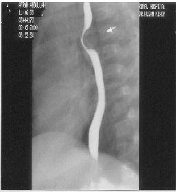
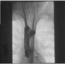
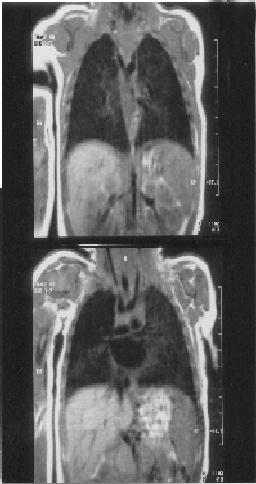
median 4.5 m, SD 6.4). To view Table I please click the link below: Preliminary investigations that led to the suspicion of the anomaly were identification of right aortic arch on chest radiograph in 7 patients and demonstration of double aortic arch on 2-Dimensional echocardiography and Doppler studies in 5 patients. Barium swallow studies documented the abnormal esophageal indentation characteristic of vascular ring (Fig. 1) in all the 16 patients. Confirmatory imaging techniques included digital subtrac-tion angiography in 7/16, cardiac catheteriza-tion and angiography in 5/16 (Fig. 2), and computerized tomography and magnetic resonance imaging of the chest (Fig. 3) in 2 patients each.
the time of surgery. In 9 patients with double aortic arch the right (posterior) arch was the smaller one and required division, while in the remaining 3 patients with double aortic arch the left anterior arch (atretic in 1 patient) was divided. 4 patients required division of the ligamentum arteriosum, and 2 among these patients had in addition excision of the aortic diverticulum (Kommerrell’s diverticulum). Immediate postoperative period was uneventful in 13 patients, who remained completely asympto-matic. Two patients developed chylothorax, which subsided on conservative management and one needed prolonged ventilation. These 3 patients continued to suffer from occasional respiratory infections. Discussion Vascular rings refer to congenital anomalies of the aortic arch and the great vessels emerging from the heart, characterized by the blood vessels completely encircling the trachea and esophagus(5). The resultant compression of the trachea and esophagus may result in symptomatic obstruction to these structures or may be completely asympto-matic. The symptoms when present are almost never cardiovascular in nature but mostly respiratory as a result of partial tracheal obstruction. Occasionally, difficulty in swallowing is a presenting symptom. Gross and Ware(6) originally described two classic true anatomic vascular rings, double aortic arch and right aortic arch with a left ligamentum arteriosum. Later two other types were described–the innominate artery compression syndrome (an abnormally distal and posterior takeoff of the innominate artery) and pulmonary artery sling (left pulmonary artery originating from the right pulmonary artery). Some of these are not in fact completely encircling and strictly speaking, may not fit the definition of vascular rings. However, in view of similar symptomatology and management approach, they are considered along with vascular rings in publications. Aberrant origin of the right subclavian artery as the most distal brachiocephalic branch from a normal left aortic arch is rare and mostly asymptomatic (7). Similarly, an aberrant left subclavian artery by itself is not pathological; however when associated with a left sided ductus arteriosus or more commonly a left sided ligamentum arteriosum, it can compress the trachea. Vascular rings are the result of abnormal development of the embryonic brachial arches. Stewart et al(8) explained all aortic arch anomalies as being the result either of abnormal regression and/or abnormal persistence of a part of the hypothetical primitive double aortic arch system. Our study of symptomatic vascular rings has shown a frequency of sub-types somewhat different from previous publications(9-17). Roberts et al(9) reported 13/30 patients with right aortic arch and aberrant left subclavian artery and only 10/30 patients with double aortic arch, while Hovarth et al(14) had equal number of patients with the two anomalies among his 22 patients. Similar frequencies have been reported by Chun et al(15) and others(9-13). Our series had a relatively higher proportion of double aortic arches. This could reflect the population studied or be merely due to the small number of patients. There are no published studies from the Arab population on vascular rings, and hence it is difficult to come to any conclusion on this aspect. Among the double aortic arches in our patients, the right (posterior) arch was dominant in the majority, a finding similar to those published by Gross and Ware(6) and Nikaidoh et al(12). Vascular rings have also been reported recently in older children and adults who had been labeled earlier as resistant asthma(18-20). The diagnostic techniques employed in patients with suspected vascular rings included plain chest radiograph, barium esophagraphy, 2-dimensional echocardio-graphy, computerized tomography, magnetic resonance angiography, digital subtraction angiography and aortic cineangio-graphy(10,21-24). Barium swallow studies were the most useful in our patients in making a firm suspicion of the vascular ring, and yielded positive results in all the 16 patients studied. However a confirmatory study for defining the anatomy was essential, and as per traditional approach, aortography had to be resorted to in the early years of the study period in 5 of our patients. Digital subtraction angiography was found to be helpful in 7 of our patients. However in the latter 5 years of the study, the diagnosis could be established by non-invasive techniques namely computed tomography in 2 patients and magnetic resonance imaging in another 2. Currently magnetic resonance imaging has been accepted as the method of choice for the delineation of the vascular and tracheal anatomy in patients suspected of having a vascular ring(22). However a recent report from Missouri(16) highlights the improve-ments in echocardiography tech-niques that allow better delineation of the anatomy, thereby limiting the use of magnetic resonance imaging to selected patients with uncommon forms of vascular rings. Gross and Ware(6) made the most significant early contributions to the operative treatment of aortic arch anomalies. Surgery of a double aortic arch involves division of the non-dominant arch. In our series, the posterior (right) arch was divided in 9 patients and the anterior (left) in 3. The remaining 4 patients required division of the ligamentum arteriosum, thereby breaking the constricting circle. Presence of an aortic diverticulum (Kommerrell’s diverticulum) necessitates resection of this diverticulum as, untreated, it may lead to residual obstruction and persistent symptomatology. Freeing up the surrounding connective tissues from the trachea, eso-phagus, and aorta further enhances the surgical procedure. Post operatively, secondary tracheomalacia may lead to incomplete resolution of symptoms, as was seen in 3 of our patients. Some surgical centers routinely practice surgical fixation of the trachea (tracheopexy) to the sternum to minimize this complication(14). Contributors: RS and PV designed the study. RS collected the data. PV and RN reviwed the literature. PV prepared the manuscript which was reviewed by RS and RN. Funding: None Competing interests: None stated.
| References
|
|
![]()