1.gif (1892 bytes)

Images in Clinical practices

Indian Pediatrics 1998; 35:1028

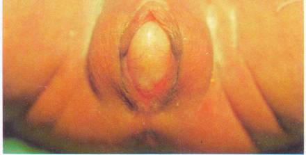
Imperforate Hymen With Hydrocolpos


This full term baby girl was born of a normal vaginal delivery. Antenatal ultra- sound had revealed a large fluid-filled cyst in the lower abdomen of the fetus. The baby presented with a palpable cystic lump in the lower abdomen. The labia were separated by a bulging yellowish white membrane (Fig. 1). On pressing the abdominal lump, the bulge in this membrane became more prominent. No other congenital anomalies were detected. Post-natal ultra- sound scan showed a fluid-filled cyst in the pelvis extending behind the bladder to the perineum. Both kidneys were normal. A diagnosis of imperforate hymen with hydrocolpos was made and the hymen was excised, draining 200ml of milky fluid from the grossly distended vagina.

 
Fig. 1. Five day old girl with imperforate hymen. Note the bulging hymen between the labia.


Imperforate hymen occurs in 1 per 2000 babies. It presents usually in the adolescent period with primary amenorrhea, cyclic abdominal pain or hematocolpos. Less often it may present in neonates or infants with an abdominal lump and an interlabial bulging membrane. The abdominal lump is due to the distended vagina (hydrocolpos) or vagina and uterus (hydrometrocolpos). Vaginal and cervical epithelium secrete mucus in response to maternal estrogens. These pool in the obstructed vagina. The distended vagina may press on the bladder outflow causing obstructive uropathy with hydroureteronephrosis or urinary infection.


Anurag Krishna,
Consultant Pediatric Surgeon,
Sir Ganga Ram Hospital,

New Delhi 110 060, India.

N .K. Arora,
Consultant Pediatrician,
Gauri Hospital,
Malka Ganj,
 Delhi
110 007, India.


Home

Past Issue

About IP

About IAP

Feedback

Links

 Author Info.

  Subscription