|
|
|
Indian Pediatr 2014;51: 909-911 |
 |
Spirometric Evaluation in Juvenile Systemic
Lupus Erythematosus
|
|
Md Mahboob Alam, *Sumantra Sarkar, *Parasar Ghosh,
Biman Ray and *Rakesh Mondal
From the Departments of Physiology and *Pediatric
Rheumatology, Institute of Postgraduate Medical Education and Research,
Kolkata, India.
Correspondence to: Dr Md Mahboob Alam, Assistant
Professor, Department of Physiology, IPGMER, 54, Alimuddin Street,
Kolkata 700 016, India.
Email: [email protected]
Received: July 05, 2014;
Initial review: July 28, 2014;
Accepted: September 20, 2014.
|
Objective: Spirometric evaluation in juvenile systemic lupus
erythematosus.
Methods: Forced vital
capacity (FVC), forced expiratory volume in 1 second (FEV1),
FEV1/FVC, forced expiratory flow between 25-75% of vital capacity
(FEF25-75%) and peak expiratory flow rate (PEFR) of 21 patients with
juvenile SLE (jSLE) were compared to controls.
Result: Reduced FVC and
FEF25-75% was found in 18 and 9 patients, respectively. All had
normal FEV1/FVC. None had respiratory complaint. When compared to
controls, patients had significantly reduced FVC [mean (SD):1.97
(0.56) vs 2.35 (0.60), P=0.002] and FEF25-75% [2.19
(0.83) vs 2.63 (0.76), P=0.028] but similar FEV1/FVC
[86.87(7.03) vs 86.72 (6.35), P=0.639].
Conclusion: jSLE patients had
significant restrictive pattern and small airway involvement.
Keyword: Pulmonary function tests,
Spirometry, Systemic lupus erythematosus.
|
|
Systemic lupus erythematosus (SLE) is a
multisystem autoimmune disorder with a broad spectrum of clinical
presentations, frequently involving dermatological, renal, neurological
and hematological systems. Pulmonary morbidities, although less
frequent, have been described in juvenile SLE (jSLE) patients [1].
Interstitial lung disease and small airway
involvement have been documented in adult SLE (aSLE) [2-4]. Evaluation
of pulmonary function abnormality is often overlooked in children, and
there is limited data in jSLE. The present study was carried out to
evaluate pulmonary function by spirometry in jSLE patients and compare
with matched controls.
Methods
This cross-sectional comparative study was carried
out from May 2013 to May 2014 in the Institute of Post-Graduate Medical
Education and Research, Kolkata, India, after obtaining clearance from
the Institutional Ethics Committee. Written consent from parents and
assent from older patients (age 12-18 yrs) were taken. All consecutive
previously diagnosed SLE patients attending the pediatric and adult
rheumatology clinic with disease onset before 16 years were included as
cases. Same number of age-sex, height- and weight-matched controls were
recruited from patients attending OPD with minor ailments and no
rheumatic or respiratory disorders. Patients under 5 years of age were
not included as they fail to understand the instruction for spirometry
[5]. Patients with chronic respiratory diseases, concurrent congenital
heart disease, congenital facial defects, and history of smoking or
surgery in the head and neck region were excluded. We performed
spirometry after recovery in those with infection, pleurisy or pleural
effusion.
Laboratory investigations included hematological and
serological investigations, examination of urine, chest X-ray,
high resolution computed tomography (HRCT) thorax, echocardiography and
ultrasonography of abdomen. SLEDAI (SLE Disease Activity Index) score
was used to evaluate disease activity [6]. Spirometry was done using
Windows-based digital spirometer (Spirowin version 2.0) after
explanation and demonstration to the subject. The nose was manually
closed by the examiner while they were asked to take maximal inspiration
and then to blow into the mouthpiece as quickly, forcefully and
maximally as possible. Forced vital capacity (FVC), Forced expiratory
volume in 1 second (FEV1), FEV1/FVC ratio, Forced expiratory flow
between 25-75% of vital capacity (FEF 25-75%)
and peak expiratory flow rate (PEFR) were noted. American Thoracic
Society (ATS) criteria for acceptability and repeatability of spirometry
were followed. Spirograms with satisfactory start and satisfactory
exhalation were considered acceptable. The spirometric procedure was
repeated until at least two acceptable spirograms showed FVC within
0.150 L of each other [7]. Maneuver with largest sum of FVC and FEV1 was
used. Patients with unacceptable spirometry and/or inadequate effort
were excluded. Global lung Initiative (GLI)-2012 equation for
‘others/mixed’ group (Quanjer) was used for calculating lower limit of
normal for FVC, FEV1 and FEF25-75%
[8].
GraphPad Prism version 5 (San Diego, CA: GraphPad
Software Inc., 2007) was used for statistical analysis. Data were
analyzed by Wilcoxon signed rank test with P value less than 0.05
considered significant.
Results
Out of 31 jSLE patients initially enrolled, five were
very sick and could not perform spirometry. Three young children failed
to perform acceptable spirometry due to problem in comprehension, while
two older children with sub-maximal effort did not meet repeatability
criteria. Of the remaining 21 patients (age 9-18 years), 20 were
females. Mean age, height and weight were 15.52 yrs, 148.0 cm and 41.81
kg, respectively. Mean duration of the disease was 2.6 years. Majority
of patients (n=13) were in remission with SLEDAI score of zero.
None had respiratory symptom at rest or with activities.
TABLE I Characteristics of the Study Poplation (N=21)
|
jSLE, Mean (SD) |
Control, Mean (SD) |
|
Age, y |
15.52 (2.93) |
15.76 (3.05) |
|
Height, cm |
148.0 (11.3) |
148.3 (9.3) |
|
Weight, kg |
41.81 (8.75) |
41.24 (7.99) |
|
#FVC, L |
1.96 (0.60) |
2.35 (0.60) |
|
*FEV1, L |
1.681 (0.43) |
2.02 (0.45) |
|
FEV1/FVC, % |
86.87 (7.03) |
86.72 (6.35) |
|
$FEF25%-75% , L/s |
2.19 (0.83) |
2.63 (0.76) |
|
PEFR, L/s |
3.56 (1.06) |
3.58 (0.94) |
|
*P <0.001; #P=0.002, $P=0.029; jSLE:
juvenile SLE patients. |
Reduced FVC and FEV1 were found in 18 (86%) patients.
FEV1/FVC ratio was normal in all. FEF 25-75%
was decreased in 9 (43%) patients. Eight patients had simultaneously
decreased FVC, FEV1 and FEF25-75%.
Table I shows the comparison of parameters
between jSLE patients and controls. FVC, FEV1 and FEF25-75%
were significantly compromised in jSLE patients. But FEV1/FVC ratio and
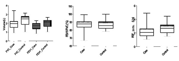
PEFR were similar to those of controls. Fig. 1 compares
median, maximum, minimum and inter-quartile range of FVC and FEV1,
FEV1/FVC and FEF25-75% of
cases and controls.
 |
|
Fig. 1 Box and whisker plot comparing FVC and FEV1
(a), FEV1/FVC (b), and FEF25-75%
(c) of cases and controls.
|
Discussion
This study, demonstrated significant restrictive
pattern and small airway involvement in jSLE. Reduced FVC, FEV1 and FEF25-75%
but similar FEV1/FVC ratio indicate restrictive pattern and small airway
involvement in jSLE patients. Small airway involvement was found in 43%
of patients and 86% had possible subclinical restrictive disease that
needs to be confirmed by Diffusing capacity for carbon monoxide (DLCO),
High resolution computed tomography (HRCT) and measurement of Total lung
capacity (TLC).
Due to lack of standardized data on spirometry, we
compared cases with age-, sex-, height- and weight-matched controls.
However, Quanjer’s equation was also used for quantitative analysis.
Exclusion of very sick children and those not fulfilling acceptability
and repeatability criteria reduced the sample size in our study. HRCT
could not be done in all children due to financial constraint. DLCO
could also not been done due to unavailability of this facility in our
institution.
Pulmonary manifestations in jSLE include pleuritis
and pleural effusion, acute lupus pneumonitis, chronic interstitial lung
disease (ILD), pulmonary hemorrhage, diaphragmatic dysfunction and
pulmonary hypertension [1,9,10]. ILD on HRCT has been documented in
nearly one-third of aSLE with no clinical symptoms [2,3]. However,
reports of ILD in pediatric population are scarce. ILD was found in 14%
of jSLE patients in one study, while another showed abnormal HRCT in 8%
but none had ILD [11,12]. Spirometry, an inexpensive and easily
available screening tool, is especially suitable for early detection of
restrictive pattern in pediatric rheumatologic diseases in a
resource-limited infrastructure [13]. Abnormal spirometry and DLCO has
been described in 20% to more than half of jSLE patients in different
studies [11,14,15]. Progressive decline in FEF 25-75%
indicating small airway disease in aSLE patients has been reported
earlier [4]. It is possible that the jSLE patients would develop
restrictive and/or obstructive lung disease at a later age.
Periodic spirometric evaluation might be a
cost-effective option to detect the subclinical pulmonary changes in
settings where DLCO or repeated HRCT cannot be carried out. Early
detection of patients ‘at risk’ of developing future pulmonary
complications by timely screening could guide the clinician for
appropriate intervention at the outset. A longitudinal multi-center
study is needed to establish the relation of pulmonary function with
duration and disease activity.
Contributors: MMA: concept and design of
study, data collection, analysis and interpretation, and manuscript
writing; SS: study concept and design, data collection and manuscript
writing; BR, PG and RM: data collection. All the authors finally
approved the manuscript. MMA will act as the guarantor.
Funding: None; Competing interests:
None stated.
|
What This Study Adds?
• There is a significant restrictive pattern
and small airway involvement on spirometry in jSLE patients.
|
References
1. Caeiro F, Michielson FM, Bernstein R, Hughes GR,
Ansell BM. Systemic lupus erythematosus in childhood. Ann Rheum Dis.
1981; 40:325-31.
2. Bankier AA, Kiener HP, Wiesmayr MN, Fleischmann D,
Kontrus M, Herold CJ, et al. Discrete lung involvement in
systemic lupus erythematosus: CT assessment. Radiology. 1995;
196:835-40.
3. Fenlon HM, Doran M, Sant SM, Breatnach E.
High-resolution chest CT in systemic lupus erythematosus. Am J
Roentgenol. 1996;166:301-7.
4. Eichacker PQ, Pinsker K, Epstein A, Schiffenbauer
J, Grayzel A. Serial pulmonary function testing in patients with
systemic lupus erythematosus. Chest. 1988;94:129-32.
5. Eigen H, Bieler H, Grant D, Christoph K, Terrill
D, Heilman DK, et al. Spirometric pulmonary function in healthy
preschool children. Am J Respir Crit Care Med. 2001;163:619-23.
6. Bombardier C, Gladman DD, Urowitz MB, Caron D,
Chang CH. Derivation of the SLEDAI. A disease activity index for lupus
patients. Arthritis Rheum. 1992;35:630-40.
7. Standardization of spirometry, 1994 Update.
American Thoracic Society. Am J Respir Crit Care Med. 1995;152:1107-36.
8. Quanjer PH, Stanojevic S, Cole TJ, Baur X, Hall
GL, Culver BH. ERS Global Lung Function Initiative. Multi-ethnic
reference values for spirometry for the 3-95-yr age range: the global
lung function 2012 equations. Eur Respir J. 2012;40:1324-34.
9. Habibi S, Saleem MA, Ramanan AV. Juvenile systemic
lupus erythematosus: Review of clinical features and management. Indian
Pediatr. 2011;48:879-87.
10. Ciftçi E, Yalçinkaya F, Ince E, Ekim M, Ileri M,
Orgerin Z. et al. Pulmonary involvement in childhood-onset
systemic lupus erythematosus: A report of five cases. Rheumatology
(Oxford). 2004;43:587-91.
11. Lilleby V1, Aaløkken TM, Johansen B, Førre Ø.
Pulmonary involvement in patients with childhood-onset systemic lupus
erythematosus. Clin Exp Rheumatol. 2006;24:203-8.
12. Beresford MW, Cleary AG, Sills JA, Couriel J,
Davidson JE. Cardio-pulmonary involvement in juvenile systemic lupus
erythematosus. Lupus. 2005;14:152-8.
13. Alam MM, Ray B, Sarkar S, Mandal O, Mondal R,
Hazra A, et al. Spirometric evaluation in juvenile idiopathic
arthritis: Data from Eastern India. Indian J Pediatr. 2014 Feb 5. [Epub
ahead of print].
14. Trapani S, Camiciottoli G, Ermini M, Castellani
W, Falcini F. Pulmonary involvement in juvenile systemic lupus
erythematosus: A study on lung function in patients asymptomatic for
respiratory disease. Lupus. 1998; 7:545-50.
15. Al-Abbad AJ, Cabral DA, Sanatani S, Sandor GG,
Seear M, Petty RE, et al. Echocardiography and pulmonary function
testing in childhood onset systemic lupus erythematosus. Lupus.
2001;10:32-7.
|
|
|
 |
|
