|
|
Letters to the Editor Indian Pediatrics 2006; 43:1007-1008 |
|||
|
Antenatal MRI in the diagnosis of Tuberous Sclerosis |
|||
|
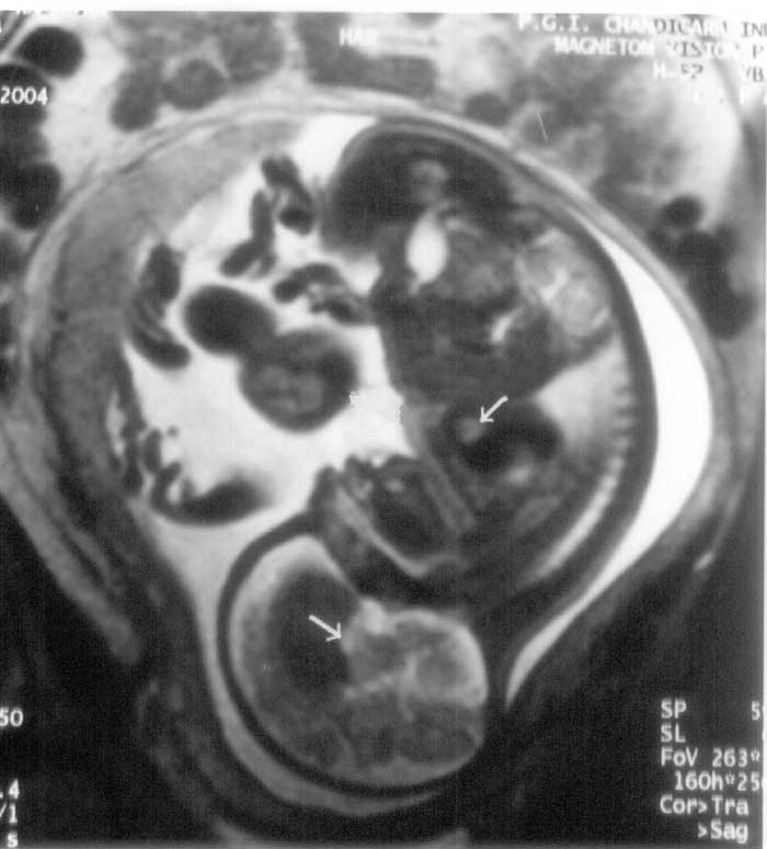
An obstetrical ultrasound examination of a 24-year-old primigravida at 22 weeks of gestation showed two hyperechogenic solid masses in ventricle of the fetal heart. Fetal MRI revealed cardiac rhabdomyoma along with a subependymal tuber in the brain causing obstructive hydrocephalus. (Fig. 1). The couple was counseled but they wished to continue the pregnancy. The baby was delivered uneventfully at term. Posnatal echocardiography confirmed the cardiac findings and MRI brain showed multiple cortical and subependymal tumors and one subependymal congenital giant cell astrocytoma (SEGA) which occluded the foramen of Monro causing hydrocephalus. The child had delayed neurodevelopment and multiple ash leaf macules in followup. He developed infantile spasms at 6 months and was treated with Vigabatrin. Ultrasound kidneys and fundus examination of the child and parents were unremarkable. Surgery for SEGA was suggested in view of the abnormally enlarging head size due of obstructive hydrocephalus which was denied by the parents in view of high risk. The neurobehavioural problems, risk of recurrence and need for antenatal screening in subsequent pregnancies were told to the parents.
Previously antenatal diagnosis of TS was done by sonographic detection of fetal cardiac rhabdomyomas(2). Eighty seven percent of fetuses with cardicac rhabdomyomas have TS, while only 50% TS have them, most of which are typically not present at the time of prenatal screening(3). Simultaneous detection of cardiac and cerebral tubers would therefore improve the diagnosis. Ultrasound again is of limited utility for fetal brain evaluation. Detection of subtle pathology like cerebral tubers is possible as early as 21 weeks by MRI due to high-spatiotemporal resolution, multiplanar capabilities and superior soft-tissue characterization. With the advent of fast, single-shot sequences, the motion of the fetus can be "frozen" and artifacts reduced(2-4). False-negative diagnoses underestimating the cerebral involvement especially if subependymal noduli and cortical tubers are <1 cm and lack of correlation with severity of neurological manifestations are the limitations of this procedure(5). Thus use of prenatal MRI in addition to sonography is a valuable tool in diagnosis and counseling for TS. Kavitha Kothur,
|
![]()