|
|
Images in Clinical Practice Indian Pediatrics 2005;42:1150-1151 |
||
|
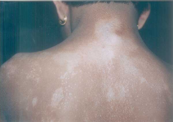
Extensive Lichen Sclerosus et Atrophicus |
||
|
LSA is an uncommon disorder of unknown etiology. Reports have suggested an association with Borrelia burgdorferi infection and also that it may be an autoimmune-related disorder. It is more common in females with M:F ratio of 1:10 and less common (2-15%) in children. LSA can present as non-genital or anogenital lesions. Non-genital lesions are usually asymptomatic and appear as ivory- or porcelain white macules, papules and plaques. Dilated pilosebaceous or sweat duct orifices filled with keratin plugs (Dells) is characteristic. Non-genital LSA is often confused with pityriasis versicolor, vitiligo, morphea or lichen simplex chronicus. Seventy five percent of children with LSA have anogenitallesions. In females, it is called kraurosis vulvae and can involve the vulva and perianal region in a ‘figure-of-eight’ pattern. It may be asymptomatic or produce severe irritation, itching and dysuria. Labial fusion, labial and clitoral atrophy and narrowing of the vaginal introitus may develop. Anogenital LSA in males termed as balanitis xerotica obliterans, produces phimosis. Treatment modalities include topical steroids, topical estrogens/testosterone, topical vitamin D3, oral steroid and PUVA therapy. Circumcision is the treatment of choice in male anogenital LSA. Vulvar LSA in prepubertal children does not predispose to neoplasia and is not an indication for surgery. Unlike adult LSA, in children, the disease is usually self-limiting and most non-genital LSA and two thirds of anogenital LSA involute around menarche. Sujay Khandpur,
|
![]()