1.gif (1892 bytes)

Images in Clinical Practice

Indian Pediatrics 2000;37: 1278.
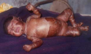
Bullous Ichthyosis


A one- month-old female child, product of non-consanguineous marriage presented with widespread areas of denuded skin, blistering and with areas of hyperkeratotic small, hard verrucous scales (Fig. 1). During subsequent 1 month, she presented with recurrent blisters which were secondarily infected. She was treated with antibiotics and Vitamin-A. After 2 months, blistering improved but hyper-keratosis persisted. Now the child is surviving and 6 months old. Histopathologic examination of skin showed epidermolytic hyperkeratosis, acanthosis and intracellular vacuolation of spinous cell layers which was consistent with bullous ichthyosis.

Bullous ichthyosis is an autosomal dominant disorder characterized by onset at birth, generalized erythroderma, recurrent bullae and severe hyperkeratosis. Verrucous hyperkeratosis becomes secondarily infected producing foul body odor. With time, blistering component diminishes while scaling compo-nent usually remains life long. Oral retinoids are moderately effective, but skeletal toxicity may occur; these should therefore be given for the shortest period at the lowest effective dose.

Fig. 1. One-month-old child with blisters, denuded skin and small warty hyperkeratotic scales.

S.K. Swain,
Specialist in Pediatrics,
U.G.P.H.C., Patkura
District Kendrapara 754 228,
Orissa, India.


Home

Past Issue

About IP

About IAP

Feedback

Links

 Author Info.

  Subscription