|
|
Original Articles Indian Pediatrics 2000;37: 1181-1187. |
||||||||
|
Non
Surgical Closure ofA atrial Septal Defect Using the Amplatzer Septal |
||||||||
|
Manuscript received: February 9,
2000; Initial review completed: March 30, 2000;
Key words: Amplatzer septal occluder, Atrial septal defect. ATRIAL septal defect is one of the commonest form of congenital heart defects seen in the general population(1). It is also the commonest congenital heart defect seen in the adult population. The defect is frequently missed in infancy and childhood because patients are asymptomatic till the third or fourth decade. However, the best results of closure of such defects are seen only when these patients are operated upon before the first decade. Early closure prevents develop-ment of arrhythmia and patient survival matches that of the general population. Till 10 years ago the only option available for closure of such defects was by open-heart surgery. Although, in most centers the mortality of ASD closure has approached 0%, the morbidity associated with operation remains an issue(3). Scar of right atrial surgical incision may play a causal role for late development of atrial arrhythmia. In this light, percutaneous closure of atrial septal defect could overcome many of the disadvantages associated with surgical closure. Non-surgical closure of ASD is under a process of evolution with the search for ideal device still underway. Since the first attempt to close ASD(4), rapid development has been made in the recent past. The plethora of devices available include that none of them may be perfect and only long term follow up will clarify whether any device will stand the test of time and match the excellent results achieved by surgery(5-11). We present here our initial experience with the Amplatzer septal occluder device in closing fossa ovalis ASD’s in children.
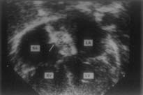
From May 1998 till date, 49 children under 12 years of age were evaluated and advised closure of ASD based on presence of defect and right ventricular volume overload as seen on 2D echocardiogram. In all patients the ASD was carefully evaluated by echocardiogram for suitability of device closure. Decision to offer device closure as an alternate method was based on size of the defect and presence of adequate margins all around the defect. In most instances these decisions were arbitrary. Thus, if it was felt that the size of the ASD size less than 15 mm with atleast 5 mm margin all around as measured on 2 dimensional echocardiogram .Borderline cases were given the option of device closure with a guarded outcome. In all cases it was carefully explained to the parents about the possibility of being referred for surgical closure in case of device failure. A total of 16 patients fulfilled our criteria for device closure. Their age ranged from 2.5 years to 11 years (mean 6.5 years). The weight ranged from 10 Kg to 42 Kg (mean 19.1 Kg). There were 7 females and 6 males. Thirty three children were not considered for device closure of the defect, the reasons for which are discussed later. The procedure was done under general anesthesia under guidance of transesophageal echocardiogram (Pediatric biplane probe, Hewlett Packard). The radial artery was cannulated for monitoring systemic pressure. The right femoral vein was cannulated and a complete right heart study was done to determine the pulmonary artery pressure and calculate shunts. A right upper pulmonary vein angiogram was done to define the location of the defect and also use this as a roadmap for subsequent procedure. The ASD was then balloon sized (Medtech Balloon Occlusion Catheter, Boston Scientific) in order to determine the stretched diameter of the defect. The size of the device chosen was 1 mm more than the stretched diameter of the defect. The Device The device (Fig. 1) is a double disc made from a nitinol wire mesh. The discs are linked together by a short waist. Each wire runs continuously from the top of the one disc, through the waist, to the end of the fiber. The use of nitinol makes the device self-expandable, since this alloy regains its preformed shape and size. In order to increase the capacity to block the defect, each disc contains a polyester patch, and the waist is filled with polyester other disc. The device, which comes in sizes varying from 4 to 40 mm for the diameter of the waist, has disc edges of 4 mm. It is screwed on its delivery cable, and fits through a long introducer sheath of variable sizes.
Devices Deployment Once the device size was determined, a long 7f to 9f mullins transeptal sheath was advanced into the left atrium. The dilator was removed leaving only the sheath in the left atrium. After taking care that all air has been removed from the sheath, the loaded device was introduced and advanced to the tip of the Mullins Sheath. Adjustments in the position of the sheath were then made so that the waist of the device, representing the central connection between the left and right atrial disc, was centered at the site of the atrial septal defect. This was done using the angiogram, showing the defect, as a road map. The sheath was then withdrawn in one continuous process, deploying the left atrial, the connecting waist and the right atrial disc in sequence. The position of the device was then checked on the transesophageal echocardiogram. If this was statisfactory the device was released and a repeate transesophageal echocardiogram done to look for residual shunt. If any problems related to device position and stability were noted before device release, then the device was retrieved into the sheath and the process of deployment repeated. Follow up An attempt was made to discharge all patients the following day. All patients were put on aspirin (5 mg/kg) and advised to continue the drug for 6 months. A repeat transthoracic echocardiogram was done the following morning for residual shunting and stability of the device. Review of clinical and echocardiographic evaluation was done at 3, 6 and 12 months.
A total of 49 patients, ages ranging between 2.5 years to 14 years, were screened for atrial septal defects. Of these 16 patients were found to be having ASD suitable for device closure. All patients had left to right shunt more than 1.8 : 1 and were associated with right ventricular volume overload. All except one patient had a single fossa ovalis defect. The one patient with multiple ASD had 3 defects in the oval fossa but closely related to each other. In this case it was thought that covering the largest defect, the device would overlap the other smaller ones. The mean ASD size was 11.1 mm (range 8-16 mm). The stretched diameter of ASD varied from 13-30 mm (mean 15 mm). The device was successfully deployed in all except three patients. Complete abolition of shunt was achieved in 5/13 cases (38%) in the immediate post deployment as seen on transesophageal study. Twenty four hours later, the transthoracic echocardiogram demonstrated complete closure in 12/13 cases (92%). The one patient with trivial persistent flow at 24 h did not show any flow at 3 months of follow up. The average fluoroscopic time was 12 minutes (Range 9 min to 20 min). Failed Attempts We were unsuccessful in three of our attempts. One patient in whom it "failed" had a stretched diameter of >30 mm and therefore the procedure was abandoned in favor of surgery. This patient had an anatomical ASD size of 16 mm at transesophageal echocardio-gram with deficient inferior rim. In two other patients the procedure was abandoned because it was felt that the device/left atrial size ratio was such that adjacent structures may be encroached upon (anatomical size 10 and 14 mm). Thirty three patients were sent for surgery directly after initial screening, their ages ranged between 1.5 to 14 years (mean age 6.1 years). ASD size ranged between 8.9-27 mm (mean 18 mm). Commonest cause for rejection was a large defect which occurred in 25/33 (76%) patients, followed by deficiency of margins seen in 4 (12%) patients, 3 (9%) children had multiple defects which were far apart and would have needed more than one device to close, and in two cases the parents opted for surgical closure despite the fact that the defect was suited for device closure. Follow up Till date, 11, 8 and 4 patients have completed 3 mo, 6 mo and 1 year follow up, respectively. No symptoms have been noticed in any of these patients. Follow up trans-thoracic echo-cardiogram revealed stable device in all. There was no residual flow and no new development of valvar regurgitation. None showed any evidence of clots on the device. Complication/Procedural Problems In two patients the device had to be pulled back into the sheath because of improper positioning of the device (release of both left and right atrial discs into the left atrium). The device was redeployed in a second attempt with success. One patient had transient ST segment elevation during withdrawal of the Mullins dilator from the left atrium.
In this small series of 16 patients, our study shows that it is feasible to close small to moderate sized atrial septal defects with the Amplatzer septal occluder with excellent immediate and short-term results. Because of ease of use of the device, the fluoroscopic time was only 12 minutes on an average. No major procedural complications or difficulties were encountered. Although there is now extensive experience with this device, we have for the first time reported its feasibility and results specifically in children. This is related to the fact that: (i) It is in this age group that closure of ASD provides maximum benefit in terms of survival and freedom from atrial arrthymias; and (ii) Device closure of atrial septal defects in children is more difficult and demands greater attention to details as compared to the adult population. The smaller size of the heart means that the larger ASD’s would be difficult to close because the device size necessary to close such defects may encroach on important adjacent cardiac structures (AV valves, pulmonary veins). In our own experience ASD’s larger than 14-15 anatomical size would be difficult to close in this age group. Another reason for failure inspite of a size, which was thought to be "adequate", was absence of adequate rims. This, in majority of instances was due to poor inferior rims (near the inferior vena cava). Thus, in 2 cases in whom the procedure was abandoned inspite of sizes which appeared suitable for device (9 mm and 14 mm), there were poor inferior margins. This resulted in stretched diameter of the defect, which precluded safe device closure. Of note, in our series, is that only 28% of children with fossa ovalis ASD were found to be suitable for consideration of device closure. This was mainly related to the size of the ASD. In patients in whom it was not attempted, the average ASD size was 18 mm. Two patients in whom a device was feasible, refused this option and opted for surgery. Another patient was not considered for device because of inadequate rims especially the inferior rim inspite of a size, which could be suitable for device closure (12 mm). Two patients had a multifenestrated ASD in which the defects were wide apart and would have required more than one device to close the interatrial defect. In the published data of device closure of ASD using the Amplatzer device, the procedure was successful in nearly 100% cases but population in all these published series was a heterogenous group of adults and children with ages ranging from 0.8-77 years (12-14). To the best of our knowledge no single study has specifically looked for the probability of use of Amplatzer device in children; hence to draw comparison from these studies may not be ideal. Referral bias may also play a role. Being a tertiary referral center only symptomatic patients may reach the center for treatment. Thus, it is possible that we might be encountering patients with larger defects. The third reason may be a more conservative approach to device treatment as a policy. Thus, in the group of patients in whom device treatment was not considered, a few patients with ASD sizes between 16-18 mm could have been candidates for device. Our decision was however based on the estimated device size that would be required in such cases (usually 1.5 times the anatomical size). Thus, in cases with sizes between 16-18 mm it was felt that the device required would encroach on adjacent structures and hence a surgical option was preferred. The cost of device is another important consideration. In most Government Institutions in our country, surgery would be a much cheaper option. However, in private institutions like ours the cost of surgery vis-a-vis device closure is competitive. Also, short hospital stay, absence of scar, no blood requirement and minimal morbidity may outweigh the disadvantage of cost. The absence of scars in the right atrium may minimize atrial arrthymias but this is an unproven hypothesis and needs to be tested on long-term follow up. Long term follow up is also necessary to document sustained benefit and absence of complications related to device placement in the heart at such a small age. The history of surgical ASD closure is 50 years old and its benefits are well proven. Meticulous follow up of patients undergoing intracardiac device especially at such young age is necessary, to demonstrate its safety vis-a-vis surgical closure. Contributors: SR was responsible for designing and monitoring the study; he will act as the guarantor for the study. AM was responsible for collecting the data and compiling the study. SS was responsible for critical analysis and drafting of the manuscript. Competing interests:
None stated.
|
![]()