|
|
|
Indian Pediatr 2014;51: 411-412 |
 |
Williams Syndrome: A Case Series
|
|
Subapriya Kandasamy, Deepti Saxena, Yougal Kishore and
Shubha R Phadke
From Department of Medical Genetics, Sanjay Gandhi
Postgraduate Institute of Medical Sciences, Lucknow, UP, India.
Correspondence to: Dr Shubha R Phadke, Professor and
Head, Department of Medical Genetics, Sanjay Gandhi Postgraduate
Institute of Medical Sciences, Lucknow, Uttar Pradesh, India.
Email:
[email protected]
|
|
Pediatricians’ awareness about malformation syndromes can help in their
timely diagnosis. Williams syndrome is a microdeletion syndrome
associated with characteristic facial features and behavioral phenotype.
Diagnosis can be confirmed by fluorescence-in-situ hybridization or
multiplex ligation probe amplification. Correct diagnosis can help in
diagnosing hypercalcemia and cardiac defects, and providing genetic
counseling to the family.
Keywords: FISH, Microdeletion, Williams
syndrome.
|
|
Williams syndrome (OMIM 194050) is caused by hemizygous
microdeletion on chromosome 7; the estimated prevalence
varies from 1: 20000 to 1:50000 live births. It is
characterized by cardiovascular disease, distinctive facies
and personality, mild intellectual disability, unique
personality, and connective tissue growth and endocrine
abnormalities. The aim of this communication is to describe
the typical facial and other diagnostic features of Williams
syndrome.
The data about the facial and other
phenotypic features of 11 patients (8 boys) of Williams
syndrome diagnosed at our center from 2007 to 2012 were
collected. The diagnosis was confirmed by
Fluorescence-in-situ- hybridization (FISH) or Multiple
Ligation Probe Amplification (MLPA).
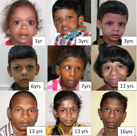
The mean age was 8 years. The presenting
complaints were developmental delay and/or intellectual
disability (in 6 children) or hyperactive behaviour (in 2
children). Fig. 1 shows facial features of 9
children with Williams syndrome. Malar hypoplasia, thick
lower lip and bulbous nasal tip were present in 10 cases;
periorbital fullness and long philtrum were seen in 8 cases.
Seven patients had bi-temporal narrowing and full cheeks,
while 6 had short nose and wide mouth. Prominent ear lobes
were seen in 5 patients. Epicanthal folds, small jaw,
malocclusion of teeth and widely spaced teeth were seen in 3
children. The cardiac anomalies (aortic stenosis, pulmonary
stenosis, ventricular septal defect and mitral valve
prolapse) were present in 5 out of 9 children in whom
echocardiography was done. Four patients had short stature
and six had microcephaly.
 |
|
Fig 1: Facial features of
Williams syndrome.
|
Jurado, et al. [1] suggested that
postnatal growth deficiency may be associated with deletion
of maternal copy of chromosome 7. One of our patients
presented at 3.5 months of age with cholestatic jaundice,
paucity of bile ducts and facial dysmorphism. The diagnosis
of Williams syndrome was made at 1 year when developmental
delay and characteristic facial features became obvious.
The facial phenotype of Williams syndrome
is characteristic and is seen in all children. The fullness
of cheeks is lost with age and face becomes long and
coarser. Cardiac malformations are commonly seen and are a
common reason for referral [2,3]. In a study from Brazil
[4], out of 23 clinically suspected
cases, 15 were confirmed to have Williams syndrome. In
addition to characteristic facial phenotype, joint laxity,
cardiac malformation and overfriendly personality help in
clinical diagnosis. Rauch, et al. [5] reported the
prevalence of William syndrome to be 1.3% in cases with
intellectual disability.
There was no infant that had
hypercalcemia in our case series. The photographs in this
series are expected to give visual impression of the facial
features that can help in diagnosis by gestalt.
Acknowledgement: Indian Council for
Medical Research.
References
1. Pérez Jurado LA, Peoples R, Kaplan P,
Hamel BC, Francke U. Molecular definition of the chromosome
7 deletion in Williams syndrome and parent-of-origin effects
on growth. Am J Hum Genet. 1996;59:781-92.
2. Patil SJ, Madhusudhan BG, Shah S,
Suresh PV. Facial phenotype at different ages and
cardiovascular malformations in children with Williams-Beuren
syndrome: a study from India. Am J Med Genet A.
2012;158A:1729-34.
3. Ferrero GB, Biamino E, Sorasio L, Banaudi
E, Peruzzi L, Forzano S, et al. Presenting phenotype
and clinical evaluation in a cohort of 22 Williams-Beuren
syndrome patients. Eur J Med Genet. 2007;50:327-37.
4. Viana MM, Stofanko M,
Gonçalves-Dornelas H, da Silva Cunha P, de Aguiar MJ.
Phenotype of Williams-Beuren syndrome in Brazilian patients:
comments on the article by Patil, et al. [2012] and
discussion of variable phenotypes in distinct populations.
Am J Med Genet A. 2013;161A:637-8.
5. Rauch A, Hoyer J, Guth S, Zweier
C, Kraus C, Becker C, et al. Diagnostic yield of
various genetic approaches in patients with unexplained
developmental delay or mental retardation. Am J Med Genet A.
2006;140:2063-74.
|
|
|
 |
|
