An 8 years-old boy, presenting with clawing of left little
and ring fingers for 2 years was referred to Department of
Dermatology for evaluation of Hansen’s disease. There was no
history of trauma or loss of sensation in affected limb or
elsewhere, but had history of skin tightening of left limb
and few patchy areas on trunk 2 years back, which resolved
within a year. The clawing of fingers followed this skin
tightening. Examination revealed no hypopigmented
hypoaesthetic lesions anywhere on the body and no peripheral
sensory loss was appreciable. The left hand was atrophic on
medial side and was notable for linear indurated lesion
extending from medial arm to palm and medial two fingers.
Overlying skin was hyperpigmented and could not be pinched
off. Similar indurated plaques were found on trunk and
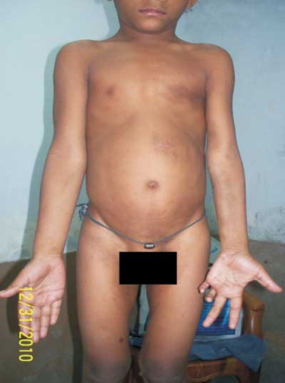
medial thighs too (Fig.1). All peripheral
nerve trunks appeared to be normal in thickness. A clinical,
diagnosis of Morphea (Linear morphea- hand lesion and
classical plaque type- trunk lesions) was made.
Histopathology was consistent with the diagnosis.
 |
|
Fig. 1 Linear indurated
plaque on left forearm (linear morphea) causing
atrophy of medial forearm and clawing of medial two
fingers. Note indurated plaques on trunk (classical
plaque type morphea).
|
Morphea is a clinically distinct
inflammatory condition affecting primarily dermis and
subcutaneous fat, which ultimately leads to scar-like
sclerosis. Classical plaque type has an asymmetric patchy
distribution on trunk; acral parts are uncommonly involved.
However, linear form preferentially affects extremities and
is much more common in children. Usually, there is no
systemic association and it is a relatively harmless
disease; however, lesions affecting extremities may result
in significant contractures and deformities. Linear lesions
are common on face too and are known as "morphoea en coup
de sabre". Differential diagnosis includes scleroderma
(symmetric acral involvement, raynaud’s phenomenon,
gastrointestinal and pulmonary function compromise),
Eosinophlic fasciitis (rapidly onset of edema of extremities
following strenuous exercise, dry river bed sign), Lupus
panniculitis (tender nodules and plaques on face, trunk and
upper arms), and lichen sclerosus (predominantly affects
genitalia, hypopigmented lesions, follicular plugging and
some evidence of hemorrhage in lesion). Diagnosis is mainly
clinical with a supportive histopathology. Early
identification and treatment is necessary to avoid lifelong
morbidities. Psoralen-UV A therapy (PUVA) and UV A1 therapy
are particularly effective. Rapidly progressing disease may
require weekly methotrexate and pulsed high dose
corticosteroid therapy. The disease has a good prognosis and
many lesions heal spontaneously in about 3-5 years. However,
older lesions may reactivate and new lesions may appear. The
major concern is irreversible fibrosis of skin and
subcutaneous tissues and around joints, which necessitates
early diagnosis and treatment.


