|
|
Images in Clinical Practice Indian Pediatrics 2006; 44:231-232 |
||
|
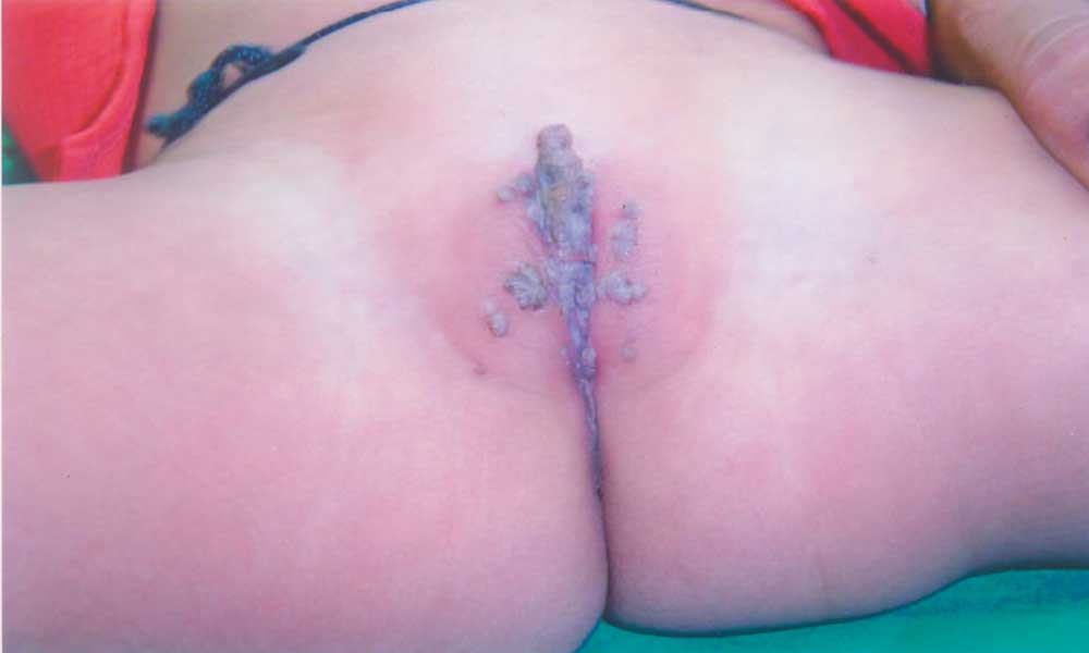
Vulval Condylomata Acuminata |
||
|
Condylomata acuminata or ‘genital warts’, caused by the human papilloma virus (HPV), are usually asymptomatic, rarely may cause pain during urination or defecation and large condylomata may even cause obstruction of the urethra. Vertical transmission of HPV can occur via in-utero exposure to amniotic fluid or transmission of HPV from the maternal genital tract. The incubation period is usually several months, the average latency being about 3 months. In children less than two years of age the virus is acquired from the mother during birth or by hetero-inoculation from warts on the hands; after two years of age, sexual abuse may be a concern. Patients diagnosed with condyloma are at increased risk for other sexually transmitted diseases like gonorrhea, Hepatitis B, Hepatitis C, syphilis and HIV. The differential diagnosis includes condylomata lata, molluscum contagiosum, skin tags and seborrheic keratosis. Condylomata lata are smooth, moist and flat lesions with surface scrapings under dark field microscopy showing typical spirochetes. Molluscum contagiosum are usually discrete, painless, flesh colored papules that classically have a central umbilication. The lesions have a predilection for the face, trunk, and extremities in children and for the groin and genitalia in adults. Seborrheic keratoses presents as one or more small, sharply defined, light brown, flat lesions of several centimeters or more, with a velvety to finely verrucous surface on normal skin and can occur on almost any site of the body, with the exception of the palms, soles and mucous membranes. Some lesions of condylomata acuminata may resolve spontaneously but most require therapy. The commonly used drugs are 25% podophyllin, 0.5 % podofilox, 50% trichloracetic acid, 5% imiquimod cream and topical intralesional and systemic alpha interferon. Other methods that can be used are cryotherapy and CO2 laser vaporization therapy under anesthesia. Jyoti Sharma, Corresondence to:
|
![]()