A 7-month female child presented with abnormal look. On examination
child had cranial asymmetry, alopecia, nevus sebaceous over left scalp,
epidermal verrucous nevus on neck, cafe-au-lait spots, multiple
pigmentary patches over abdomen, bilateral corneal hazziness, skin tags
at outer canthus and dermoid of both eyes since birth (Fig. 1).
Child’s weight was 5.8 kg., length 64 cm, upper segment 39 cm, head
circumference 43.5 cm with normal developmental age. Routine hematology,
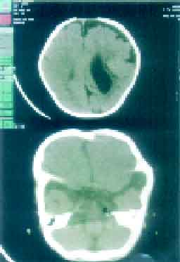
B-Scan of eyes, ECG, ECHO and abdominal USG were normal. CT scan head
revealed-lipoma (HU value –40 to –60) at left cerebello pontine angle,
10 mm × 7 mm × 5 mm size, with dilated left ventricle, cortical atrophy
(Fig. 2). A diagnosis of encephalocraniocutaneous lipomatosis was
made.
 |
 |
| Fig. 1.
Clinical Photograph showing the features of
encephalocraniocutaneous lipomatosis. |
Fig.2. CT
scan depicts dilated left lateral ventricle with lipoma
(arrowhead) at left cerebello-pontine angle. |
Encephalocraniocutaneous lipomatosis is a rare
congenital neurocutaneous disease. It is characterized by unilateral
lipomatous hamartoma of scalp, eyelid and outer globe of eye,
ipsilateral porencephalic cyst, intracranial lipoma, cortical atrophy,
cranial asymmetry, developmental delay and mental retardation. The
clinical picture may vary from patient to patient. Mental status varies
from totally normal to severe mental retardationr seizure may be
associated in some cases. Bilateral cutaneous and visceral involvement
is also rarely reported. The pathogenesis remains unknown. Dysgenesia of
the cephalic neural crest and the anterior neural tube is a most widely
accepted theory.
Rajiv Rathoriya,
Jyotsna Shrivastava,
Department of Pediatrics,
Gandhi Medical College,
Bhopal, Madhya Pradesh, India.
E-mail:
[email protected]
