Cushing’s disease is bilateral adreno-cortical
hyperplasia secondary to excessive secretion of ACTH by the pituitary
gland and is very rare in children(1,2). Very few cases have been
reported in Indian and world literature(3). Cushing’s syndrome in
pediatric patients is usually caused by adrenal tumors(4), as opposed to
this pituitary tumors causing Cushing’s disease are extremely rare under
the age of 7 years and more so in infancy(3). We report a case of an
11-month-o1d child with Cushing’s disease caused by a Pituitary macroadenoma.
Case Report
An 11-month-old female child, first issue of
non-consanguineous marriage was referred for obesity. On examination the
patient was 77cms long (97th centile Agarwal charts) and weighed 14 Kgs
(above 97th centile Agarwal charts). She had generalized obesity, blood
pressure was 140/110 mm of Hg using a mercury sphygmomanometer, moon
facies, edema, hirsuitism, clitoral hypertrophy and dark pigmentation.
Within 3 weeks patient started showing visual inattention though the
fundoscopic examination was normal.
Investigations showed a raised midnight
Adrenocorticotrophic hormone (ACTH) (122 pgm/ml), serum Prolactin (100
ngm/ml), and plasma Cortisol (88 mcg/dL) and post prandial glucose of 72
mg%. Thyroid stimulating hormone was 3U/ml (0.4-5 U/mL), Thyroxine was
10 microg/dL (5-12 microgm/dL), Tri- iodothyronine was 120 ng/dL(70-190
ng/dL), serum sodium was 141 (136-152 meq/lit), serum potassium was 2.3
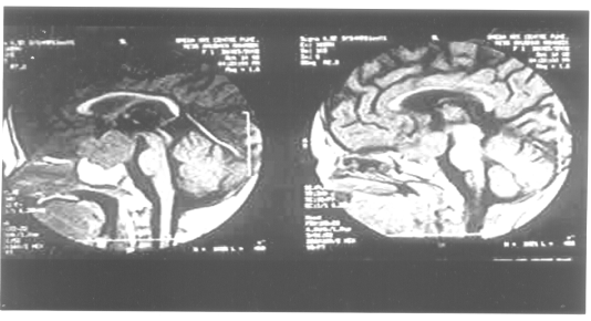
(3.5- 5.6 meq/lit). An MRI of the brain showed the presence of a large
pituitary macroadenoma measuring 3 × 2.5 × 2.5 cm (Fig. 1)
elevating the optic chiasma. No calcification was seen. Both the adrenal
glands were well visualized on CT scan of the abdomen suggesting
bilateral adrenal hyperplasia. On the basis of physical examination,
endocrine and neuro-radiologic assessment, diagnosis of Cushing’s
disease was made. Since neurosurgical experience in Pituitary
macroadenomas in infancy is limited it was decided to manage the patient
medically with Cyproheptidine and Bromocriptine. After three weeks of
medical therapy patient developed visual inattention and hence immediate
surgery was planned. As sphenoid sinuses are not developed at this age
trans-sphenoidal surgery was not possible. Trans-cranial surgery was
performed. The tumour was successfully removed however, despite careful
monitoring and Pediatric ICU care patient succumbed due to severe
electrolyte imbalance in the immediate post-operative period.
 |
|
Fig. 1. MRI showing large pituitory
macroadenoma . |
Discussion
The term Cushing’s disease is currently used for
condition in which bilateral adrenocortical hyperplasia is secondary to
excessive secretion of ACTH by the pituitary gland. The adrenals may be
slightly or greatly enlarged. Reviews of literature reveal that
Cushing’s disease is extremely rare in infancy(3). Pituitary Cushing’s
may be caused by a pituitary adenoma though macro-adenomas are rarely
found in children (5). Most adenomas found in children are under one cm
and do not extend outside the pituitary fossa. When the pituitary
adenoma extends outside the fossa it grows upwards into the suprasellar
recess and compresses the optic chiasma as happened in our patient.
The signs and symptoms of Cushing’s disease are due
to cortisol excess and to pressure symptoms caused by the pituitary
adenoma. Frequent clinical findings include weight gain, truncal
obesity, straie, hyper-tension, glucose intolerance and infections.
Progressive obesity is often the first symptom as was seen in our
patient.
The evaluation of patients with suspected Cushing’s
disease and syndrome requires an understanding of the proper use and
limitations of the tests commonly included in the diagnostic work-up.
Estimation of urinary free cortisol, study of circadian rhythm, low dose
and high dose Dexamethasone suppression test may be required for a
definitive diagnosis and for locating the tumour. For neuroimaging
Magnetic Resonance Imaging with enhancement is the diagnostic
investigation of choice. Surgical excision of an ACTH-producing
pituitary tumour is the optimal therapy for Cushing’s disease. However,
medical therapy may have either a primary or adjunctive role if the
patient cannot safely undergo surgery. The medications work through
three broad mechanisms. "Neuromodulatory" compounds modulate
corticotropin (ACTH) release from a pituitary tumor e.g.,
Bromocriptine and Cyproheptidine, steroidogenesis inhibitors reduce
cortisol levels by adrenolytic activity e.g., Mitotane,
Metyrapone, Ketoconazole, and Aminoglutethimide, glucocorticoid anta-gonists
block cortisol action at its receptor e.g., Ketoconazole(6). In
the past, attempts have been made to manage Cushing’s disease by medical
therapy viz., Cyproheptidine and Bromocriptine without much long
term success(3). Cyproheptadine and bromo-criptine have been reported to
be therapeutic in suppressing ACTH levels in Cushing’s disease.
Surgery is the treatment of choice in all patients
with pituitary tumors and the trans- sphenoidal removal of the tumor is
the treatment of choice. However in infants as the sphenoidal cells are
not formed and the pnematization is only complete by 4 years(5), this
option is not available. Death in the immediate post-operative period as
was seen in our patient is also reported in a similar case(7).
This extremely rare case demonstrates that though a
pituitary macroadenoma is extremely rare at this age it should still be
considered as a cause for Cushing’s syndrome in infancy although the
commonest cause at this age is adrenal tumors.
Contributors: VVK and JRN carried out the
clinical workup. A VK reviewed the literature and drafted the
manuscript. VVK supervised drafting of the paper and will act as
guarantor for the paper.
Funding : HCJMRI, Jehangir Hospital, Pune.
Competing interests: None stated.
