|
|
Case Reports Indian Pediatrics 2002; 39:299-304 |
||
|
Incomplete Beckwith-Wiedemann Syndrome in a Child with Orbital Rhabdomyosarcoma |
||
|
V. Thavaraj Anitha Sethi* L.S. Arya
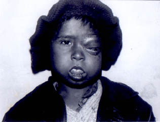
Beckwith-Wiedemann syndrome (BWS) was first recognized as a syndrome by Beckwith in 1963, when he reported autopsy findings on three unrelated children with omphalocele, macroglossia, cytomegaly of adrenal cortex, renal medullary hyperplasia and hyperplastic visceromegaly(1). In 1964 Wiedemann published an article on the same syndrome in three siblings with omphalocele, macrosmia and neonatal hypoglycemia(2). The incidence of BWS has been estimated to be 1:13700 live births(3). The increased risk of tumor formation in BWS patients is estimated to be 7.5%(4) and the risk is further increased to 10% if hemihypertrophy is present(4). It has been suggested that BWS can be inherited by the homozygous state of an autosomal recessive mutation(5). We describe a rare case report of partial expression of BWS associated with orbital rhabdomyosarcoma (RMS). Case Report A 5-year-old boy presented to the Pediatric Oncology Clinic at All India Institute of Medical Sciences, New Delhi with the chief complaints of swelling of the left eye for the last 3 months. He was apparently alright 3 months ago when the parents noticed swelling of the left eye which gradually increased in size (Fig. 1). There was no history of white reflex, trauma to the eye, redness, watering, decreased vision or deviation of the eyes. There is a history of the child born with a large tongue. However, the large tongue did not give rise to any respiratory or feeding difficulties in the early neonatal period. There was no family history of cancer or BWS. The child is a product of a non consanguineous marriage, a full term and normal delivered baby. He was not a large weight baby. Even though the mother remembers that he was reddish pink in color at birth. He developed normally except that the speech was delayed and he had articulation problem.
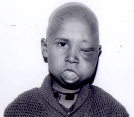
On examination he was average built child. His weight was 15.0 kg (10th percentile) and height was 108.0 cm (between 25th and 50th percentile). Left eye was proptotic. The right eye was normal. The preauricular and anterior cervical group of lymphnodes were not palpable. The tongue was large and with mouth open all the time. He was unable to close the mouth. There was no hypertrophy of any limbs of the body. There was no hepatosplenomegaly. Both the scrotal sacs were empty due to undescended testes. The ultrasonography of the eyes revealed left eye structurally normal. There was a mass outside the eyeball. The contrast enhanced computerized tomography (CECT) of the head and orbit revealed a left extraconal mass suggesting an embryonal RMS and there was no extension of the tumor into the central nervous system or the adjoining structures. The ultrasonography of the abdomen and scrotum to locate both the testes was done. Bilateral testes were not located in the scrotal sacs or in the inguinal canal. There was no hydrocele. The retroperitoneum was also normal. Liver, kidney, gall bladder, pancreas were normal in echotexure. There was no masses or lymph nodes seen in the abdomen. The cytogenetics studies were not carried out in this patient. The metastatic work up including bone marrow biopsy with touch smears and the cerebrospinal fluid cytopathology for spill of tumor cells were negative. The histopathological examination of the biopsy from left eye revealed, malignant mesenchymal tumor compatible with embryo-nal RMS. The child was started on chemo-therapy each cycle consisted of Vincristine 1.5 mg/m2 IV (day 1, 15, 28, 42), Actinomycin-D 20 µg/kg IV day 1-5 and cyclophosphamide 10 mg/kg/day 1-5 and Adriamycin 40 mg/m2 on day 28. The cycles were repeated every 9 weeks. A total of 5 cycles were given. The child also received radical external beam radiotherapy to the left orbit. He received 55 Gy in 23 fractions in 4.3 weeks. The child is on follow up (Fig. 2) and in complete remission for the last 20 months. Discussion DeBaun et al. reported 0.027 cancer/person/year to be the average annual incidence of cancer in the first four years of life in 183 children with BWS, who were followed up longitudinally for 482 person years(6). There is increased risk of develop-ment of both benign and malignant tumors in BWS. Incomplete forms of the syndrome are recognized and share the same potential risk for development of neoplasia as the complete forms(6). Wiedemann observed 29 cases of BWS developing 32 neoplasms with tumor rate of 7.5%(4).
Fig. 2. Rhabdomyosarcoma regressed completely with chemotherapy and in remission. The malignant tumors reported in full blown BWS are nephroblastoma adrenal cortical carcinoma, hepatoblastoma, glio-blastoma of brain stem, neuroblastoma, mesoblastic nephroma and RMS(4,6-10). The malignant neoplasms associated with incomplete forms of BWS reported in literature are nephroblastoma, adrenal cortical carcinoma, hepatoblastoma and hepato-cellular carcinoma(7). Wilm’s tumor is most frequent embyonal tumor followed by adrenal cortical carcinoma and hepato-blastoma(9,11,12). The overall risk occur-rence of Wilm’s tumor in BWS is 3-5% of the cases(13). In a series of neoplasms associated with BWS, Wilm’s tumor was seen in 63% of cases followed by two cases of hepato-blastoma, one case each of adrenal cotical tumor and RMS of pelvis arising from bladder(8). RMS is rarely associated with BWS. There are only few reports in the literature(7,8,11-15). This case report of RMS is from the left orbit. There is only one case of report of RMS of orbit associated with BWS(7). The other cases of RMS with BWS are from urinary bladder (8,14,15). One case of embryonal RMS from pelvis associated with an isolated hemihypertrophy has also been reported(16). Exomphalos, macroglossia and gigantism are considered the characteristic diagnostic triad in BWS and is also referred as EMG-Syndrome(17). The following abnormalities are seen in various combinations in patients with BWS: macroglossia, defects of closure of abdominal wall (omphalocele, umbilical hernia, diastasis recti), facial flame nevus, visceromegaly, natal and post natal gigantism, advanced bone age, ear lobe abnormalities, microcephaly, somatic asymmetry, absence of gonads, clitoral hypertrophy, diaphragmatic abnormalities (eventration, hernia) and muscular hypertrophy, mental retardation, neonatal hypoglycemia, secondary poly- cythemia and combined immunodeficiency syndrome and hypertension in adolescence is also seen(7). BWS has a variable clinical expression and clinical features may occur in various combinations(7). This patient had macroglossia and bilaterally absent gonads (testes). There were no other congenitial abnormalities. He was of normal intelligence and possibly had a secondary polycythemia in the neonatal period due to the history given by the mother that he was reddish pink in color. Macro-glossia is the most frequent manifestation of BWS and may cause feeding difficulties and speech delay secondary to articulation problem. Macroglossia is seen in 98% of BWS(18). Surgical tongue reduction is performed in upto 50% of cases usually at 2-3 years of age. The relative risk of cancer associated with macroglossia is 1.7 in BWS(6). The differential diagnosis of macro-glossia may be divided into true and relative macroglossia(19). The causes of the true macroglossia includes vascular malforma- tion (hemangioma, lymphangioma), muscular enlargement (hemihypertrophy BWS), sys-temic disorders (mucopolysaccharidoses and amyloid) and tumors. The relative macro-glossia could be due to systemic disorders like Down’s syndrome, patient having an abnormal small oral cavity. Congenital anomalies were identified in 37 of 115 (32%) autopsies of children and adolescents who had died from RMS(20). The authors had reported various congenital anomalies of the central nervous system, cardiovascular system and genitourinary system. But they had not found macroglossia and bilateral absence of gonads in these 115 cases of children who had died of RMS(20). Familial studies indicate linkage of the BWS locus to the marker chromosome 11p 15.5(21), and the insulin like growth factor (IGF)-II gene located at this locus has been suggested as a candidate for the BWS gene(22). Increased levels of IGF-II mRNA have been detected in numerous human tumors often associated with the BWS such as nephroblastoma, neuroblastoma, rhabdo-myosarcoma and adrenal cortical carci-nomas(23). In conclusion, we describe a patient with a partial expression of Beckwith-Wiedemann syndrome associated with an unilateral orbital RMS presented at the age of 4 years and 9 months, Chemoradiotherapy was used to treat RMS and he is in complete remission for the last 20 months. Contributors: VT and LSA treated the case and prepared manuscript. AS interpreted the biopsy for diagnosis. VT will act as the guarantor for the manu-script. Funding: None. Competing interests: None stated.
| ||
| References | ||
|
![]()