|
|
Letters to the Editor Indian Pediatrics 2004; 41:627-628 |
|||
|
Splenic Abscess Treated with Percutaneous Aspiration |
|||
|
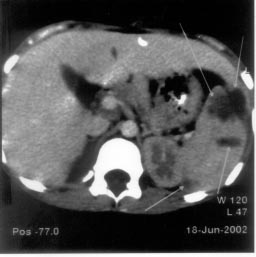
A 10-year-old girl presented with high grade intermittent fever for 2 weeks and left lower chest pain for 4 days. There was no history of trauma. On examination she was febrile and had tenderness in left lower half of chest with diminished breath sounds in the same region. Hepatomegaly (4 cm) and tender splenomegaly (3 cm) were present. Rest of the examination was unremarkable. Investiga-tions showed normal blood counts and a high ESR (60 mm fall in 1 hour). USG abdomen showed splenomegaly with a focal hypo-echoic area measuring 15 mm in diameter. Widal test showed normal titers. Blood culture was sterile. Sickling test was negative. Chest x-ray showed elevated left hemi- diaphragm. Lung parenchyma and costo-phrenic angles were normal. A presumptive diagnosis of splenic abscess was made and the child was started on Inj. ciprofloxacin and gentamicin. One week of antibiotic therapy did not result in significant improvement. A CT scan abdomen done at this stage showed splenomegaly with multiple well defined irregular hypodense non-enhancing areas, the largest one measuring 2.8 cm (Fig. I), which was drained percuta-neously. Pus culture was sterile. AFB stain and Gram stain were negative. There was a significant improve-ment in the condition of the child following drainage of the abscess. She became afebrile with regression of hepatospleno-megaly. Antibiotic were continued for two more weeks. USG abdomen repeated twice during this period showed regression in the size of abscess in the first scan and a normal spleen with no hypoechoic areas in the next scan.
Splenic abscess may develop after generalized infection, hematological disorder and trauma. The commonest cause is hematogenous seeding of the spleen from an infective focus elsewhere in the body. Infecting microorganisms include gram positive bacteria, mycobacteria, fungi and anerobes(3). USG detects large abscesses easily, but may miss the small abscesses. CT scan remains the gold standard for definitive diagnosis(4). CT is also useful for diagnostic/therapeutic aspiration. Recent trends in the management of a splenic abscess employ techniques like percutaneous catheter drainage and fine needle aspiration of the abscess(3,5). In our case, needle aspiration with use of antibiotics resulted in cure. In children spleen should be preserved as far as possible. Aggressive surgical procedures like splenectomy should be reserved for the non-responders. T.S. Raghu Raman,
|
![]()