Acute renal failure (ARF) is frequent in newborns and
infants. Clinical conditions causing hypovolemia, hypoxemia and
hypotension in newborns and infants may lead to renal insufficiency. Few
leading causes are perinatal anoxia, ischemia and sepsis. ARF accounts
for 8-24% of all NICU admissions. Intrinsic renal failure accounts for
11% of all admissions of which intra-renal obstruction is rare(1,2). We
report two infants with intra-tubular obstruction presenting as ARF.
Case Reports
Case 1
A 3-month-old girl, first living child born to third
degree consanguineous parents was referred for management of ARF with
oliguria, which was noticed during evaluation of acute gastroenteritis.
The patientís antenatal, perinatal, birth and developmental history was
unremarkable except for repeated episodes of vomiting. On examination
the infant was dehydrated, tachypneic, acidotic with a short systolic
murmur over the left precordium medial to the mid-clavicular line with
normal blood pressure. There was no renal or bladder mass.
Investigations showed blood urea level of 333 mg/dL,
creatinine 9.1 mg/dL, bicarbonate 12 mEq/L and hemoglobin 6 g/dL.
Ultra-sonogram of abdomen showed both kidneys of 5.1 cm size with
uniformly hyperechoic renal parenchyma and global nephro-calcinosis.
Echocardiography showed a small ventricular septal defect. The patient
was started on intravenous fluids with sodium bicarbonate and peritoneal
dialysis. Ultra-sound guided renal biopsy, done five days after
admission, showed 16 glomeruli with some degree of immaturity, shrinkage
of the tuft and corona of prominent visceral epithelial cells. The
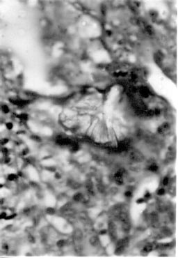
cellularity and basement membrane thickness appeared normal. Striking
features were seen in the tubules, many of which were filled with
crystalline deposits, obstructing the lumina completely. In some areas
the deposits were seen within the interstitium extending from the
tubular wall. Patchy infiltrates of lymphocytes were seen in the
interstitium. Blood vessels appeared mildly thickened and the biopsy
picture was suggestive of oxalosis (Fig. 1). 24-hr urine oxalate
excretion was 14 mg for the 5 kg child (normal <2 mg/kg/day). The
patient was started on treatment with oral pyridoxine 100 mg daily and
discharged on request with serum creatinine level of 4.5 mg/dL.
 |
|
Fig. 1. Kidney biopsy showing oxlate
crystals in the tubules. |
Case 2
A 3-month-old girl born to non-consanguineous parents
was referred for acute gastroenteritis with dehydration, hurried
breathing and oliguria. Blood pressure was normal. Evaluation showed a
serum creatinine of 7.9 mg/dL, urea 162 mg/dL and bicarbonate 8 mEq/L.
Ultrasonogram showed global nephrocalcinosis. She was treated with
intravenous fluids and peritoneal dialysis. An ultrasound guided renal
biopsy showed 17-18 glomeruli with mild increase in mesangial
cellularity and increased mesangial matrix. Many tubules showed
destruction of the lining epithelium and contained crystalline material,
consistent with oxalate crystals, which was birefringent under polarized
light. The interstitium showed patchy aggregates of lymphocytes and
mononuclear cells. The patient was treated with oral pyridoxine 100 mg
daily and discharged on request with a serum creatinine level of 3.2 mg/dL.
Discussion
Oxalosis is characterized by elevated levels of
oxalic acid. Oxalosis can be primary or secondary. Primary oxalosis is
of two types, type I and type II. The childhood and young adult form of
oxalosis present with history, signs and symptoms of urolithiaisis
whereas infantile type of primary oxalosis present with renal failure.
Hence sibling with oxalosis, neonate with ARF and an infant with
echogenic kidney should be taken as clues for early diagnosis(3,4). It
is imperative to screen a sibling of a child with oxalosis antenatally,
with the help of amniocentesis, chorionic villus sampling and fetal
liver biopsy(5). Liver biopsy tissue showing deficiency of the enzyme
alanine glyoxylate aminiotransferase is the diagnostic investigation.
Preemptive liver transplantation has a better therapeutic strategy than
combined liver and kidney transplantation after the onset of renal
failure, to improve the outlook of these patients( 6).
Difficulties in the diagnosis of oxalosis in infancy
are many. The normal range of urinary and plasma oxalate, glycolate,
glyoxylate and glycerate in infants are not known. Serum oxalate level
tends to be higher in children with oxalosis and renal failure when
compared to children with same status of renal failure without oxalosis.
Diagnosis of oxaluria may be complicated by the presence of renal
failure as urine oxalate excretion may decrease and even fall within the
normal range when renal failure becomes more advanced.
Oxalosis should be considered in the differential
diagnosis of intrinsic renal failure in infancy especially when on
clinical evaluation there are no signs or symptoms other than those,
which can be explained by renal failure. An ultrasonogram must be done
in every child with renal failure(7). Documentation of cortical/global
nephro-calcinosis in an infant warrants further careful evaluation. Thus
ARF when associated with cortical nephrocalcinosis is a strong pointer
for oxalosis especially with an insignificant past history. Exact
diagnosis is important since it has consequence concerning genetic
counseling and treatment. Hence in the absence of a liver biopsy, which
is confirmatory, an ultrasonogram will suggest and renal biopsy will
confirm oxalosis. Dialysis and transplantation are hard to justify in
infantile primary oxalosis at present.
Acknowledgment
We thank the Department of Histo-pathology, Apollo
Hospitals, Chennai for their help in histopathological examination of
kidney biopsy specimens.
Contributors: NP, MVK and BRN were involved in
care of the patient and preparation of the manuscript. BRN will act as
the guarantor for the paper.
Funding: None.
Competing interests: None stated.
